Filtered by author: Nicole Quain Clear Filter

AAWC Shares A Common Mission

The Association for the Advancement of Wound Care (AAWC) is a society of wound care providers from a variety of clinical specialties, backgrounds, and levels of expertise. Each member brings their unique perspective, experiences, and insights that are extremely valuable. The AAWC thrives off of this collective knowledge and welcomes all clinicians, as well as wound care industry professionals.

Our diversity is our strength. It allows our members to contribute ideas that otherwise may never have been considered or to give testimony about an experience that others may have never encountered. The AAWC is comprised of nurses, physicians, advanced practice providers, podiatrists, occupational therapists, physical therapists, professors, Ph.D. scientists, and researchers, among many other professions. Beyond these differences, our members represent various races, religions, genders, sexual orientations, and socioeconomic statuses, among other diversities.

We are all united by a common mission: to advance the care of patients with and at risk for wounds.

Together, we rise to the challenges and collaborate to identify scientifically-based practices that support decisions that will ensure the optimal outcome for our patients. The AAWC emphasizes collaborative efforts for the betterment of our members, patients, healthcare practices, and communities.

We invite you to help further our cause and join, rejoin, or renew your membership with our association. Visit our member benefits and options to learn more. 

The AAWC is here for you. We invite you to join us and contribute your insights to our association and advance the field of wound care, for a better, and more inclusive, tomorrow.

Sincerely,
The AAWC President and Communications Committee

AAWC President Ruth Bryant, Ph.D., MS, RN, CWOCN

Amanda Blackmon, PT, DPT, CWS, CSWS, CMTPT

Blair Flinn, MHA

Jenneffer Pulapaka, DPM, ACFAS, CWSP, DABMSP, FACCWS

Maria Goddard, MD, CWS, FAPWCA

Nicole Rodriguez, LPN, CWCA, WCC

Rose Hamm, PT, DPT, CWS, FACCWS

Saralyn McDade, BSN, RN,WOCN, CWCN, CSWS

Victoria Nalls, GNP-BC, CWS, ACHPN, Nurse Practitioner, Capital Caring

The Association for the Advancement of Wound Care & The Alliance of Wound Care Stakeholders


AAWC is a member of The Alliance of Wound Care Stakeholders, a nonprofit, multidisciplinary trade association of medical specialty societies and clinical associations whose mission is to promote quality care and access to products and services for people with wounds through effective advocacy and educational outreach in the regulatory, legislative and public arenas.

As both “watchdog” and go-to resource, the Alliance impacts clinical and procedural policies. In an effort to remove or reduce a barrier to treatment, the Alliance requested a ‘clarification’ of the non-covered surgical dressing benefit. To support the request, the Alliance conducted a teleconference with the Medical Directors from the Durable Medical Equipment Medicare Administrative Contractors (DMEMACs) and the Pricing, Data Analysis, and Coding (PDAC) Contractor. The Alliance afforded these medical directors an opportunity to interact with clinical wound care “experts”, including two AAWC Board members, who offered actual examples of patients who benefit from treatment that the proposed clarification would support.

Information sourced from the Alliance website on July 5, 2020. Please visit www.woundcarestakeholders.org for more information.

Wound Care in Crisis: Navigating unique challenges and new environments

As I watch various media cover the health care response to the COVID-19, I am reminded of my past experiences with disaster relief. Specifically, I am reminded of the opening of a Federal Medical Shelter (FMS) for special-needs evacuees during Hurricane Rita and Ike. At that time, I was deployed as an officer with the United States Public Health Service to work with fellow officers, as well as volunteers.

As a wound care nurse, I did not fully realize the impact our specialty can have during crisis response situations. After all, it had been many years since I worked as a nurse at the bedside, or in any of the intensive care units and the ER, so I did not feel that I was clinically competent to work in various situations. I was proven wrong every day, and I quickly learned that my thoughts of inadequacy were unfounded.

In September 2005, Hurricane Rita and Ike hit the southern United States. Special-needs evacuees were being sent to shelters from nursing homes, hospice, homes where the family had assisted in their care, facilities that no longer had electricity and running water, and facilities that had been physically devastated by the natural disaster. This population brought pressure injuries, feeding tubes, chronic venous insufficiency ulcers, and various other wound care complications, but their care could not stop just because they had been removed from their normal environment.

Many of the volunteers at the shelter came from various disciplines and professional backgrounds. Most did not have the clinical skills that wound care nurses possess. During my time at the FMS, I was able to put together a wound care team of physical therapists, occupational therapists, and nurses who had similar clinical wound care experience. We all understood our specific scope of practice and standards of care. Using a team approach, we were able to perform dressing changes, clinical assessments, and documentation that could not have been accomplished without our collective expertise. We also performed discharge skin assessments and created wound care plans for evacuees as they were transferred from the FMS to receiving facilities, in collaboration with case managers and social workers.

At FMS I quickly learned who my logistics and supply officers were and found out what supplies were readily available to perform wound care. I contacted product representatives and asked for any useful supplies that they could provide. Those few weeks spent in the FMS revealed my worth as a wound care specialist. I realized that wound care clinicians have a unique set of skills, no matter what comprises our clinical environment.

The biggest help came from the local wound care nurses that lived near the FMS. Their hospital had been impacted by physical damage and had no electricity. Despite their hardships, they brought the FMS extra supplies and helped when they could. I remember their generosity well, and I plan to reciprocate their kindness if the need ever arises in my community.

These are the lessons I learned when providing disaster relief… 

  • Keep yourself healthy and hydrated. Take breaks as many times as your schedule permits since work hours will be long and physically draining.
  • Be mindful of your mental health. You are exposed full-time to a lot of suffering, loss, and grieving. Take the time to find the “good” in the situations you encounter and de-stress whenever time allows.
  • Remember that you are not in it alone and that teamwork is extremely important.

One of the most impactful lessons that I have learned during my disaster response experiences is that wound care clinicians bring a unique perspective and clinical expertise to every situation. We are privileged to serve our communities, colleagues, and patients – especially when it is most needed in crises.

About the Author

Candice Cotton, RN, MSN, CWON, is a registered nurse who earned her BSN from the University of Alabama in Huntsville, Alabama, and her MSN from the University of Phoenix, in Albuquerque, New Mexico. Cotton retired from the United States Public Health Service as a commissioned officer in 2011. She has worked in various clinical settings and served her last 16 years in PHS as a certified wound/ostomy nurse assigned to Navajo Area Indian Health Service, in Gallup, New Mexico. Currently, Cotton is working part-time as a wound/ostomy specialist at Huntsville Health Care Systems in their outpatient wound/ostomy clinic in Huntsville, Alabama.

A Cut Above: The Struggles of a Woundcare Provider During a Pandemic

As healthcare practitioners, COVID-19 has changed the way we practice in ways we never saw coming. It seems like overnight our world was turned upside down and with this, the way that we practice healthcare. As a physical therapist, I am very social and the personal time I spend with each of my patients is important to their overall care. While I have not given up this aspect of care by limiting exposure, I have had to change what it looks like. I have found ways to show my patients I care and to advocate for them from a distance. While COVID-19 has led to multiple struggles, it does not need to prevent us from continuing to provide quality care.

While I have always viewed it important that my patients have a thorough understanding of what they should be doing at home, it has become the most important part of my care plan. I have had to teach patients who do not feel comfortable changing their dressings how to do so to limit their exposure in the clinic. More than ever, I have become a cheerleader for these patients, encouraging them that they are capable of doing things they never thought they would have to, such as taking a lead role in their dressing management. I cannot stress enough the value of well-written instructions and pictures to help these patients out. E-visits have proved to be very beneficial as well to allow me to walk the patient through the process; though many of my patients who are getting wound care are not tech-savvy, so again I am doing a lot of coaching to help get them set-up. I go into each Zoom meeting with a mindset that I am going to need to be patient because sometimes half of the battle is getting the patient signed in. This battle is worth it because it means a lot to the patient to see your face and know that you took the extra effort to help them out. By getting the patient to a point where they are comfortable with being a part of the wound care team, we can ease some of their worries during this stressful time.

We have heard and experienced time and time again the difficulty with finding PPE to use with our patients. What has proved just as difficult for me is managing the PPE during wound care procedures. First of all, I wear glasses and with every breath I take, they fog up. It makes it very difficult to debride a wound when you cannot see it! First, I tried keeping my glasses a little farther from my face, which stopped the fog but led to my glasses falling off. I have researched multiple methods to assist with this, many of which seemed as if they would either decrease the effectiveness of my mask or leave a film on my glasses. While not 100% effective, the soapy water technique worked best for me;
“Immediately before wearing a face mask, wash the spectacles with soapy water and shake off the excess. Then, let the spectacles air dry or gently dry off the lenses with soft tissue before putting them back on. Now the spectacle lenses should not mist up when the face mask is worn,” said the study in the Annals of The Royal College of Surgeons of England.

Along with the fogging problem, allergy season is hitting my region. I am constantly fighting the urge to remove my mask and itch my nose. Unfortunately, there is no magic trick for that one, so I am just going to have to deal with it. At least I am no longer battling a double struggle with foggy glasses and allergies.

For me, the biggest challenge of all during the Pandemic has been getting in contact with other healthcare providers. We are all well aware of the importance of a team with wound care. As a physical therapist, I am unable to perform a culture without a script or prescribe an antibiotic and I must have a good physician by my side. As clinicians have decreased their hours in the clinic, it has made it very difficult to get anything done promptly. Let us use my patient, we will call her Sally, to drive this point home. Sally first started seeing me about a month ago for her venous insufficiency ulcers. During our time together, she met with a vascular surgeon and it was decided that surgery would be necessary. Unfortunately, this surgery was deemed “elective” and canceled. Due to pain, Sally could not tolerate any compression, which we all know is a vital aspect of healing these ulcers.

Our goal went from healing the wounds to wound maintenance until she could have surgery. Within the next two weeks, the wound size continued to increase, with worsening pain and erythema. It became clear that we needed a culture but I was unable to reach the dermatologist who had referred her due to his limited schedule. Her primary care physician only works one day a week, but we decided we would try to work with him to address the possible infection. Despite multiple phone calls it took over a week to get a culture for Sally. Our current system was not created to function well under a limited exposure schedule where providers are rarely checking their messages, and this led to Sally suffering. I am happy to say that, despite this struggle, there is a silver lining. Her infection was eventually addressed and the decision to postpone her surgery was reversed.

The last few weeks have been a whirlwind for the healthcare community. COVID-19 has forced us to change how we perform our jobs and how we treat our patients. While it has been a struggle, I do believe we will come out with a stronger skill set and better ability to think outside the box. I look forward to the day when we can all say, “We made it through this, and we are stronger for it!”.

About the Author 

Amanda Blackmon, PT, DPT, CWS, CSWS, is the Director of Premier Therapy Services at Premier Bone & Joint Centers. She grew up in Laramie and completed her undergraduate studies at the University of Wyoming, receiving a B.S. in Kinesiology and Health Promotion. She attended physical therapy school at Creighton University where she received her doctorate degree. Amanda is certified in dry needling, wounds, orthotics, myofascial trigger point therapy, sportsmetrics, custom splinting, neurological rehab and TMJ. Amanda is an active member of the AAWC and serves on the AAWC Communications Committee. 

Face Protection Protocol and Visuals for COVID-19 Frontline Staff

Disclaimer: This document is intended for educational and informational purposes only. It does not constitute medical advice. Follow institutional policies, manufacturer recommendations and principles of sound clinical judgment. It is the responsibility of each healthcare professional to verify with their institutional infection control team that any measures taken to prevent or manage PPE-related skin injuries do not interfere with the efficacy of the PPE, nor are in conflict with any workplace policy. Individuals may require a repeat mask fit testing to ensure prevention and management efforts do not interfere with PPE efficacy. Clinicians should seal check N95 respirator masks before beginning patient care.


I am a wound care nurse practitioner at a level one trauma center in Washington DC. As my hospital started ramping up new protocols and policies for COVID-19 patients, one of the things that we looked at was proning patients without the use of specialty beds. We updated policies around protecting bony prominences and the face. We quickly started gathering supplies for this expected surge in treatment. I also was spending quite a bit of time wearing N95 masks. I had forgotten how tightly they fit on the face. The tightness is for a reason, you need the seal to prevent any bacteria or viruses from being aerosolized into your airways, however, we were looking at situations where it was highly likely that staff would need to wear these masks for hours at a time, if not their entire shift. I did a simple Google search of “nurse’s faces coronavirus” and was horrified at the skin breakdown and bruising that I saw.

I quickly realized that this was something that I could work on to help protect the faces of all of the frontline staff including nurses, physicians, respiratory therapists, and the environmental service workers who are cleaning the rooms. My goal is to prevent them from suffering a painful wound while they are trying to protect themselves. I considered my options and thought of different dressings that I had on hand in my facility and things that we could use to protect people's faces and their ears. I was noticing that people were also having a breakdown on the top of their ears and behind their ears.

With a national call to wear face masks, you have probably seen the creative things that people are doing with masks such as using buttons or paperclips to hook the elastics/ties so that it doesn't rub against the ear. I tested different products to include hydrocolloid dressings, bordered and non-bordered foam dressings, and silicone interfaces.

The critical thing that drove my decision making was that those who wear a mask need to be able to pass an N95 fit test. I spoke with my chief medical officer about my plan and he quickly approved me to continue working on this protocol. I met that day with infection control, the director of the ICU, and the manager of respiratory therapy. I underwent an extensive fit test with the protocol that I developed, and fortunately, passed. I was fit tested for approximately 20 minutes and then one of my coworkers who is a male (and has facial hair) was also fit tested for about 20 minutes and he passed. I then passed the protocol to our hospital leadership and everyone agreed that this was important, and we should roll this out immediately.

These are the protocol and visuals I created for my hospital:

There are a few key points that I would like to make:

  1.  Any protocol or dressing products that you choose to use should be fit tested to ensure that there is an appropriate seal. I recommend that you involve stakeholders in leadership such as infection control, your respiratory therapy services, and chief medical as well as chief nursing officers.
  2. When you are doing a fit test or donning an N95 mask with preventive dressings in place, it is critically important that you walk your fingers away from the bridge of your nose down the metal piece by millimeters as shown in the PDF. It is simply not enough to pinch at the top at the bridge of your nose and ensure that you have a good seal.
  3. I believe that there are plenty of products which could meet the need. In this instance, we used what we had readily available.
  4. One key consideration that I used when I was developing this protocol was the interface with the skin, as I worried that people would have adhesive skin reactions or dermatitis from using certain products. This consideration was based on my personal experience.
  5. Do not apply lotion to the face right before you are placing a dressing on your face as this could also impact the seal. The face should be clean and dry before placing any of these products on the face. Moisture and hydration are very important, and I urge you to continue to moisturize your faces after you remove your masks and no longer need to wear them for that day.

I hope that you consider using something like this in your facility to protect our frontline staff. Please feel free to use it and amend it as you see fit.

Stay safe everyone! You are all heroes.

For additional resources, visit the AAWC Wound Care and COVID-19 webpage. 

A new paradigm for diagnosis and treatment of edemas: The Endothelial Glycocalyx Layer

Introduction

Although edema can result from a variety of conditions, medications or other contributing factors, it is now understood that all edema is lymphedema through a spectrum of lymphatic insufficiency.1 This article will highlight the latest evidence supporting this paradigm shift by looking at the new understanding of hemodynamics at the Endothelial Glycocalyx Layer, and the associated links between the lymphatic and integumentary systems. Further, it will explain how this information is relevant to clinical practice to help you differentially diagnose and manage lower extremity edema.

New lymphedema paradigm

One of the most significant recent changes regarding lymphedema is a more refined explanation of fluid hemodynamics impacting our historical understanding of Starling’s Law.  Previously, it was thought that 90% of fluid moving from the blood to the interstitium was reabsorbed back into the venous end of the capillary, yet the lymphatic system was only responsible for managing 10% of the fluid load. The new paradigm of the Endothelial Glycocalyx Layer (EGL) as the gatekeeper of fluid filtration from blood capillaries explains how there is only diminishing net fluid filtration across the blood capillary bed and no reabsorption at the venous end; 100% of all interstitial fluid is reabsorbed by the lymphatic capillaries alone during homeostasis. 2-3

Acting as a complex molecular sieve, the EGL precisely regulates fluid and protein movement through the capillary wall into the tissues.4-6 Conversely, the EGL also prevents movement of proteins and fluid back into the venous side of the capillaries, even when interstitial hydrostatic pressure is increased, or tissue oncotic pressures remain higher within the blood capillaries. Thus, all fluid and proteins exiting the blood capillaries must be removed from the interstitium by the lymphatic capillaries alone.  This has led to the new understanding that all edemas are on a lymphedema continuum and represent relative lymphatic insufficiency or failure. 1,7 The system is either temporarily overwhelmed (transient lymphedema/dynamic insufficiency) or the system is abnormally developed, damaged or permanently impaired leading to the disease of chronic lymphedema (mechanical lymphatic failure).

Lymphedema pathophysiology

The lymphatic system is analogous to the body’s sewer or recycling system.  It is responsible for maintaining fluid homeostasis by managing interstitial fluid and mobilizing waste products (proteins, senescent cells, macromolecules, etc.).  The lymphatic system is also tasked with the absorption and transportation of lipids and fatty acids to the circulatory system, and transporting antigens, antigen-presenting cells and other immune cells to the lymph nodes where adaptive immunity is stimulated.  Collectively, all components within the fluid transported by the lymphatic system are called the “lymphatic load”.9

Pathophysiologically, chronic lymphatic dysfunction or failure presents unique changes affecting the integumentary system.  When the lymphatic load is not readily reabsorbed by the lymphatic system from the interstitial tissues, a pathohistological state of chronic inflammation results. Free radicals trapped in the tissues denature proteins and oxidize cell membranes attracting monocytes to the area that differentiate into macrophages. These macrophages take in proteins through pinocytosis, which activates the macrophages to release cytokines. This, in turn, activates fibroblasts, which are stimulated to produce excess collagen.8,9 Excess collagen formation causes connective tissue proliferation and fibrosis resulting in the thickened, fibrotic skin and wart-like projections (papillomatosis and verrucous) commonly seen with chronic lymphedema.10 Additionally, other fibroblasts differentiate into adipocytes.9 If treatment is not implemented, the chronic inflammatory process persists and the clinical presentation eventually can result in enlargement of the body part, thickened and fibrotic dermal and subcutaneous tissues, and other significant integumentary changes.11

Disorders of the lymph system, whether systemic (macro-lymphedema) or localized (micro-lymphedema), produce cutaneous regions susceptible to infection, inflammation and carcinogenesis.10,12-13 The inter-relationship of the lymphatic and integumentary systems is starting to become more readily appreciated as a functional lymphatic system is essential to an organism’s overall health given its role in fluid homeostasis, removal of cellular debris and mediating immunity and inflammation.14 The chronic inflammation resulting from lymphedema creates a region of cutaneous immune deficiency or a localized skin barrier failure.  The associated abnormalities are called lymphostatic dermopathy, which is the failure of the skin as an immune organ.10,12-13 Because of this, alterations in skin integrity, recurrent infections (commonly cellulitis), venous dermatitis, diminished wound healing, various dermatological conditions, and even skin malignancies become more prevalent highlighting the inter-connectedness of the lymphatic and integumentary systems.10,12-13 Impairment or dysfunction in one system leads to associated complications in the other.

Edema/lymphedema examination

In combination with a comprehensive medical history and medical workup, a physical exam of the edema and its characteristics is essential. This exam should include the following simple tests. First, observe the extremity for subtle changes in contours indicative of edema and note any associated skin changes. Listen to the patient. Often, they will feel a heaviness or fullness in their limb before noticeable clinical signs. Next, cradle and gently palpate the limb circumferentially as you slowly move up from the toes or fingers to the groin or shoulder. Manually identify areas that feel full, taught, edematous or fibrotic. Next, perform the Bjork Bow Tie Test in these areas.

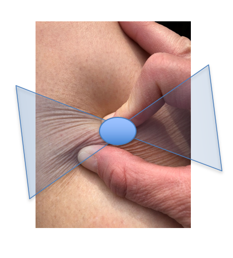
The Bjork Bow Tie Test is an expanded version of the Stemmer’s Test.15,16 The Stemmer’s Test is performed by pinching the skin at the base of the second toe or middle finger.17 If the skin can be lifted and pinched, the test is negative. A negative test does not exclude lymphedema. Thickened skin with fibrotic soft tissue changes will not lift and approximate when pinched and thus produce a positive test. The Stemmer’s Test is never falsely positive and leads to a definitive diagnosis of lymphedema.9,17 However, a limitation of the Stemmer’s Test is that, by definition, it is to be performed on the toe or finger only. And, based on the new paradigms of the microcirculation and definitions of lymphedema, all swelling can technically be diagnosed as lymphedema. The question is, what type of treatment intervention should follow? Thus, the Bjork Bow Tie Test was developed to expand the application of a Stemmer’s type test to any area of the body, as well as identify soft tissue changes, such as fibrotic tissue, that may warrant interventions to help remodel the soft tissue back to a normalized state.15,16

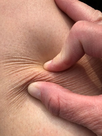
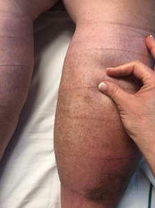
Unlike past perceptions of lymphedema presenting as gross swelling, marked fibrotic soft tissue changes or disfigurement, the subtle dermal changes are most important in early diagnosis and recommendations for care. To perform the Bjork Bow Tie Test, in one maneuver, gently pinch, roll and twist the skin between the thumb and pointer finger, noting the quality of tissue texture and thickness. Healthy skin can be lifted and pinched, should feel slippery between the layers when rolled, and produce a “bow-tie” of wrinkles when twisted. Skin that tests positive will be thickened, less pliable, less able to be pinched and lifted, more difficult to twist, and produce limited “Bow Tie” of wrinkles. A positive test indicates signs of thickening and fibrotic tissue texture changes as a result of lymphedema-induced chronic inflammation. Figure 1 demonstrates how to perform the Bjork Bow Tie Test and the difference between a negative and a positive test.

In addition to tissue texture changes, circumferences or girth can be measured using a cloth tape measure. For more precise measurement, new scanning technologies are emerging that scan a limb within minutes using an iPhone or iPad interface, create a 3-D avatar of the limb, as well as calculate volumes.18 In addition, breast and truncal edema can be measured and quantified using a hand-held, pocket-sized moisture meter that objectively measures the moisture content of affected versus unaffected areas.19 This can assist in objectively identifying areas of lymphedema that are often subtle or subclinical.

Lymphedema diagnosis

According to the new microcirculation paradigm, all patients presenting with swelling do in fact have lymphedema to some degree. The lymphatic system may be overwhelmed resulting in transient lymphedema (i.e., ankle sprain, CHF) or the system may be damaged leading to the disease of lymphedema.  Even when no swelling is present, risk factors may be identified leading to a Stage 0 lymphedema diagnosis of subclinical lymphedema.17 Table 1 lists many of the risk factors and co-morbidities contributing to lymphedema. Early identification and intervention are key, and the contributing factors and underlying comorbidities must be addressed through comprehensive medical management in order to achieve the best patient outcomes.  Utilizing the components of Complete Decongestive Therapy in the context of the patient’s entire medical picture will allow for safe and effective treatment.  The authors are now referring to this management as Lymphatic and Integumentary Rehabilitation.

In the United States, the most common cause of lymphedema in the upper extremities is breast cancer20 and in the lower extremities, chronic venous disease is the most important predictor for the development of lymphedema.21 With respect to obesity, lymphatic dysfunction can occur with a body mass index greater than 50, and lymphedema may be universal in patients with a BMI greater than 6018. Various other contributing co-morbidities and co-factors may lead to lymphedema, and most clinical presentations of lymphedema are resulting from a combination of approximately seven co-morbidities.22 Data from the Canadian LIMPRINT study showed that the most common underlying cause of lymphedema in an outpatient wound clinic was venous disease, 72% of patients had a history of cellulitis, and almost 40% had an open wound.23

Conclusion

Edema is a common and prevalent condition presenting clinically from mild to severe.  Looking at the presentation and quality of the edema, its characteristics (turgor, texture, pitting/non-pitting), associated integumentary findings, combined with a comprehensive medical review of the patient will help in determining where on the lymphedema continuum the patient resides.  Many edemas are transient, due to lymphatic insufficiency, which should fully resolve with proper medical management once the underlying cause or contributing factors have been identified and modified.  For chronic lymphedema due to lymphatic failure, managing the underlying medical issues in combination with Complete Decongestive Therapy will help the patient manage this life-long disease. 

Table 1: Lymphedema risk factors

Adapted from Framework L. Best practice for the management of lymphoedema. International consensus. London: MEP Ltd. 2006:3-52.

  • Chronic venous insufficiency
  • Post-thrombotic syndrome
  • Vein stripping or harvesting
  • Surgery (i.e. revascularization, TKA, THA, abdominal surgery, hysterectomy)
  • Decreased mobility (aging, CVA, TBI, SCI, immobilization, etc.)
  • Obesity
  • Congestive heart failure
  • Chronic kidney disease
  • Trauma
  • Scars
  • Burns
  • Lymph node dissection or removal
  • Radiation
  • Chronic wounds
  • Recurrent cellulitis
  • Congenital malformation of lymphatic vasculature
  • Tumors obstructing lymphatics
  • Travel or living in Lymphatic Filariasis endemic areas
  • The prolonged dependency of the limb or other body parts
  • Hyperthyroidism
  • Medications with edema as a side effect
  • Chronic skin disorders and inflammation
  • Arteriovenous shunt

Figure 1: Bjork Bow Tie Test

Negative Bjork Bow Tie Test
Used with permission, courtesy Robyn Bjork

Positive Bjork Bow Tie Test
Used with permission, courtesy Suzie Ehmann

“Bow Tie” of wrinkles in Negative Test
Used with permission, courtesy Robyn Bjork

Heather Hettrick PT, Ph.D., CWS, CLT-LANA, CLWT, CORE is a Professor in the Physical Therapy Program at Nova Southeastern University in Florida. As a physical therapist, her expertise resides in integumentary dysfunction where she holds four board certifications/credentials. She is actively involved with numerous professional organizations, speaks on the national and international circuit, and is faculty and Director of Wound Education at the International Lymphedema & Wound Training Institute.

Robyn Bjork, MPT, CWS, CLT-LANA, CLWT is Founder and President of the International Lymphedema & Wound Training Institute. She is a Physical Therapist who holds multiple board certifications in wound and edema/lymphedema management. Bjork is a featured speaker at national & international conferences and is dedicated to the advancement of Lymphatic & Integumentary Rehabilitation.

References

  1. Mortimer PS, Rockson SG. New developments in clinical aspects of lymphatic disease. The Journal of clinical investigation. 2014 Mar 3;124(3):915-21.
  2. Bjork R, and Hettrick H. Endothelial glycocalyx layer and interdependence of lymphatic and integumentary systems. Wounds International. 2018; Vol 9 Issue 2:50-55.
  3. Levick JR, Michel CC. Microvascular fluid exchange and the revised Starling principle. Cardiovascular research. 2010 Mar 3;87(2):198-210.
  4. Reitsma, S., Slaaf, D.W., Vink, H., Van Zandvoort, M.A. and Oude Egbrink, M.G., 2007. The endothelial glycocalyx: composition, functions, and visualization. Pflügers Archiv-European Journal of Physiology454(3), pp.345-359.
  5. Weinbaum, S., Tarbell, J.M. and Damiano, E.R., 2007. The structure and function of the endothelial glycocalyx layer. Annu. Rev. Biomed. Eng.9, pp.121-167.
  6. Woodcock, T.E. and Woodcock, T.M., 2012. Revised Starling equation and the glycocalyx model of transvascular fluid exchange: an improved paradigm for prescribing intravenous fluid therapy. British journal of anaesthesia108(3), pp.384-394.
  7. Bjork R, and Hettrick H. Emerging Paradigms Integrating the Lymphatic and Integumentary Systems: Clinical Implications. Wound Care and Hyberbaric Medicine. 2018; Vol 9 Issue 2:15-21.
  8. Scelsi R, Scelsi L, Cortinovis R, Poggi P. Morphological changes of dermal blood and lymphatic vessels in chronic venous insufficiency of the leg. International angiology: a journal of the International Union of Angiology.  1994; 13(4):308-311.
  9. Földi M, Földi E, Strößenreuther R, Kubik S, editors. Földi's textbook of lymphology: for physicians and lymphedema therapists. Elsevier Health Sciences; 2012 Feb 21.
  10. Carlson A.  Lymphedema and subclinical lymphostasis (microlymphedema) facilitate cutaneous infection, inflammatory dermatoses, and neoplasia:  A locus minoris resistentiae.  Clinics in Dermatology. 2014;32: 599-615.
  11. Shoman H, Ellahham S.  Lymphedema:  a mini-review on the pathophysiology, diagnosis, and treatment.  Vasc Dis Ther.  2017;2(3):1-2. DOI: 10.15761/VDT.1000124
  12. Ruocco E, Puca RV, Brunetti G, et al.  Lymphedematous areas:  Privileged sites for tumors, infections, and immune disorders.  Int J Dermatol. 2007;46:662.
  13. Ruocco V, Schwartz RA, Ruocco E.  Lymphedema:  An immunologically vulnerable site for the development of neoplasms. J Am Acad Dermatol. 2002;47:124-127.
  14. Ridner SH. Pathophysiology of lymphedema. Semin Oncol Nurs. 2013;29:4-11.
  15. Bjork R, Ehmann S. STRIDE Professional Guide to Compression Garment Selection for the Lower Extremity. Journal of Wound Care. 2019 Jun 1;28(Sup6a):1-44.
  16. Bjork R, Hettrick H. Emerging Paradigms Integrating the Lymphatic and Integumentary Systems: Clinical Implications.  Wound Care and Hyperbaric Medicine. 2018;9(2):17-23.
  17. Framework L. Best practice for the management of lymphoedema. International consensus. London: MEP Ltd. 2006:3-52.
  18. Greene A. Diagnosis and Management of Obesity-Inducted Lymphedema. Plastic Recon Surgery. 2016 July;138(1):111e-118e.
  19. Greenhowe J, Stephen C, McClymont L, Munnoch DA. Breast oedema following free flap breast reconstruction. The Breast. 2017 Aug 1;34:73-6.
  20. Rockson S.  Diseases of the Lymphatic Circulation in Vascular Medicine: A Companion to Braunwald’s Heart Disease, the 2nd Edition. Elsevier;2013:697-708.
  21. Mortimer P, Rockson S. New developments in clinical aspects of lymphatic disease. J Clin Invest. 2014 Mar;124(3):915-21.
  22. Wang W, Keast DH. Prevalence and characteristics of lymphoedema at a wound-care clinic. Journal of wound care. 2016 Apr 1;25(Sup4): S11-5.
  23. Keast DH, Moffatt C, Janmohammad J. Lymphedema IMpact and PRevalence INTernational (LIMPRINT) study: the Canadian data. Lymphatic Research and Biology. 2019 Mar 10.

The Pressure Ulcer Journey – From Prevention and Detection to Case Management

Pressure Ulcers can be complex, mystifying, and cumbersome for even the most experienced wound care clinician. This March, the Association for the Advancement of Wound Care (AAWC) is hosting it’s third annual Pressure Ulcer Summit (PrU), March 27-28, 2020, at the Sheraton Atlanta Hotel, to expand clinicians a wealth of knowledge surrounding pressure ulcers and the implementation science that goes into whole patient care.

The AAWC Pressure Ulcer Summit is uniquely designed to create a journey from the initial diagnosis of a pressure ulcer to navigating insurance and its complete treatment. Each educational segment builds off the prior segment’s topic and discussion. Over two days, AAWC will take clinicians through the four main stages of identifying, treating, healing and supporting patients with pressure ulcers. In this article, AAWC will provide a closer look at the educational sessions that will shed light on pressure ulcers and the expertise needed to treat the whole patient, not just the hole in the patient.

Pressure Ulcer Prevention & Detection

Pressure ulcers can often seem inevitable. The Agency for Healthcare Research & Quality (AHRQ) estimates more than 2.5 million individuals in the US develop pressure ulcers annually. Pressure ulcer prevention continues to be a challenge for even the best clinicians. The first educational segment of PrU will break down the myth surrounding pressure ulcer prevention. Through expert-led sessions, clinicians will learn about the causation and physiology behind pressure ulcers, the pathophysiologic processes, and factors that contribute to pressure-induced tissue damage and take a deeper dive into learning how to identify the conditions needed to create a pressure ulcer.

Through a better understanding of the Pressure Ulcer Detection Tool (PUDT), the three comorbid conditions that are often confused with induced tissue damage, mechanisms that contribute to tissue loss, and techniques for early pressure ulcer detection, clinicians will be able to quickly assess and accurately identify a pressure ulcer.

Once a patient is diagnosed with pressure ulcer…a clinician must face how to proceed with treatment.

Focusing on the Whole Patient, Not the Hole in the Patient

By the afternoon the PrU journey will advance beyond prevention and early detection to diagnosis. Now clinicians must use their clinical thinking and reasoning skills to solve their way through the challenges that come along with pressure ulcer treatment.

In the second segment of PrU, clinicians will explore what constitutes skin failure and develop techniques to approach difficult conversations including, debridement, wound care, pain control, antibiotics, and repositioning. Expert faculty will share their perspectives on the gaps that exist in the current method used to define skin failure and strategies for non-opioid management of painful wounds.

As experienced wound care clinicians know, pressure ulcer cases are unique to each patient, and by looking at the whole picture, clinicians can use a multidisciplinary team approach to select the best treatment strategies that will enhance a specific patient’s comfort and quality of life. While at PrU, clinicians will have the opportunity to meet other professionals in the field and develop their resource network to call upon when faced with a challenge in the real world.

With a treatment plan in place, a clinician’s next step is to select a support surface and figure out how to get it paid for.

Choosing the Right Support Surface & Getting it Paid For

In preparation for PrU day two, clinicians have explored pressure ulcer prevention, detection, and learned how to select the optimal treatment plan based on a whole-patient approach. Now faculty will tackle the complicated and often-times frustrating task of choosing support surfaces and securing payment.

Patients with pressure ulcers and persons at risk require assistance with protecting their at-risk areas. Support surfaces provide a means to decrease the compressive forces these areas experience during daily activities. These activities may include lying in bed, sitting up in a wheelchair or on the toilet and even walking. There is a broad range of support surfaces to choose from and each surface has benefits and challenges. Choosing the optimal support surface requires understanding and incorporating a broad range of physiologic and functional needs into the assessment.

To make the support surface selection and payment decision clearer, experienced faculty will identify the categories of support surfaces in detail, explain the insurance requirements needed to get payments for support surfaces, teach clinicians how to conduct an assessment that considers a patient’s physiologic and functional needs and share the staffing and clinic structure needed to set clinicians, and their patients, up for success.

With the right support surface in place and payment secured, clinicians turn their attention to case coordination and management until treatment is completed. 

Case Coordination & Management

When treating a patient with a pressure ulcer, clinicians must be empowered with the resources, tools, facility, and staff to support each stage of treatment. At the final educational segment of PrU, clinical practitioners, managers, patients, and case management experts will share strategies for successfully navigating challenging insurance, provider and other obstacles to ensure optimal outcomes.

To ensure optimal outcomes, faculty will educate clinicians on, the clinical data that is needed to set up a clinic, the variety of pressure-relieving equipment that is available, identifying equipment and support needs, prioritizing data that is available, and how to use patient engagement strategies to enable a full recovery.

After the AAWC PrU journey, clinicians will walk away with real-world knowledge, strategies, and techniques to support and treat patients with complex pressure ulcers. For more information on the AAWC Pressure Ulcer Summit (PrU) and the half-day Keeping Calm Under Pressure: How to Address Pressure Ulcers in the Post-Acute Setting and Beyond Pre-Conference on March 26th, visit www.aawcsummits.com.

A Fresh Take on Comprehensive Patient Care in Wound Healing

“Before you heal someone, ask him if he’s willing to give up the things that make him sick.” — Hippocrates

As caregivers, where should we begin when we look at comprehensive patient care? We begin with the whole patient, the whole person. We must develop a robust personal mission statement around patient care, which helps determine one's direction and focus on future goals. It aligns strategies and can give a person/team a way to reflect, evaluate, and improve outcomes in practice, such as, “I believe every patient needs a physician who will be an advocate to help them regain their health.” Strong mission statements can produce profound changes and benefits for your patients, but to do so involves viewing the patient as a whole person, not perseverating on the wound care dynamics in disunion.

The impact of conventional medicine's "sick-care" focus on our medical systems have repercussions. Is conventional medicine the best in care? It is the practice of acute treatment targeting pathogens, traumatic events, or the long-term practice for controlling disease and/or it's risk factors that reduce or degrade long-term primary and secondary outcomes. It remains a disease-focused approach with patients as recipients of “sick-care.” Patients need only comply with treatment and are not required to make significant changes or commitments to their health. The providers are responsible for the care and outcomes, while the patient is a passive receiver of services.

The "patient-centric" approach can be achieved with the use of a lifestyle medicine approach to treat, reverse, and prevent all too prevalent lifestyle-related chronic disease AND improve health literacy. Lifestyle medicine involves the use of evidence-based lifestyle therapeutic approaches, such as a predominantly whole food plant-based diet, regular physical activity, adequate sleep, stress management, avoidance of risky substance use, and other non-drug modalities to treat, reverse, and prevent all too prevalent lifestyle-related chronic disease. Most wound care patients do not experience discreetly limited events.

Acute and impaired wound healing impact our population differently. In the United States, chronic wounds affect approximately 6.5 million patients, with the majority of the chronic wounds beginning as minor traumatic injuries [see reference 1]. When wound healing is impaired, it is usually not because a single factor failed, but rather, numerously neglected and ignored comorbidities lead to an impaired healing process. A literature review of common wound care treatment options underscores to lack of whole-person care when clear evidence-based options are available and provide improved wound healing. There are several areas a provider and team can influence change. Build a referral base, create action plans, or write prescriptions that address nutritional assessments, increase exercise, improve sleep, and influence positive emotions. Below are a few ways to implement well-care with patients.

  1. Talk to patients about their diet. Nutrition is a complex field that wound care has taken a pro-active approach over the last decade. However, recently, new studies have evaluated the impact of dietary protein in patients with chronic kidney disease (CKD) and end-stage renal disease (ESRD). Shockingly, in the 2018 United States Renal Data System report, only 10% of individuals who were classified by laboratory measurements as having Stage 1-3 CKD, were aware of their kidney disease. CKD patients include poorly controlled diabetes mellitus, peripheral vascular disease, chronic venous insufficiency, and aging.

    Wound care providers should address the co-morbidity of kidney disease, protein intake, and chronic wound regimens with dietary intervention. Providers unfamiliar with the complexity of nutrition can refer patients to a skilled practitioner, such as a registered dietician, to monitor protein levels, and help address problems that arise when providers increase dietary proteins to promote wound healing. The problem emerges when an increase in protein intake increases both glomerular filtration and renal tubular acid excretion; therefore, intensifying renal injury in patients. Dietary protein intake varies for adult dialysis patients is ∼1.2 g/kg body weight of proteins per day, as opposed to non-dialysis patients, the daily protein is 0.6-0.75 g/kg.

    Lower protein intake, as reported by Klahr et al in the study, The effects of dietary protein restriction and blood-pressure control on the progression of chronic renal disease. Modification of Diet in Renal Disease Study Group, slows the progression of CKD. Another study by Kamper and Strandgaard, Long-Term Effects of High-Protein Diets on Renal Function, noted,  "daily red meat consumption over years may increase CKD risk, whereas white meat and dairy proteins appear to have no such effect, and fruit and vegetable proteins may be renal protective."

    Finally, increasing protein intake in older patients during critical illness may lead to azotemia due to decreased renal functional reserve and possibly augment the propensity towards worsened renal function and worsened clinical outcomes reported Dickerson in Nitrogen Balance and Protein Requirements for Critically Ill Older Patients.

  2. Vascular disease wound patients benefit from decreasing free fats and increasing fruit and vegetables that are rich in flavonoids to improve vascular health; reduce endothelial dysfunction, inflammation, and oxidative stress, according to Macready et al in the study Flavonoid-rich fruit and vegetables improve microvascular reactivity and inflammatory status in men at risk of cardiovascular disease--FLAVURS: a randomized controlled trial.

    Sustained dietary nitrate ingestion, via the nitric oxide (NO) cascade, improves vascular function in hypercholesterolemic patients: a randomized, double-blind, placebo-controlled study by Velmurugan et al. titled Dietary nitrate improves vascular function in patients with hypercholesterolemia: a randomized, double-blind, placebo-controlled study. These changes are associated with alterations in the oral microbiome and, in particular, nitrate-reducing organisms, which give rise in circulating levels of nitrite derived from dietary nitrate. Acute dietary nitrate load causes a marked reduction in BP, reduces platelet activation, and protects against endothelial injury. There is emergent evidence supporting the idea that CVD risk can be reduced by a dietary pattern that provides more plant sources of protein compared to animal-based dietary protein. It is a multifaceted reason that links the amino acid content of particular foods with the gut microbiome to the overall wellness of a patient. And yet, there remains a little emphasis on patient nutrition or even physical activity.

  3. Physical inactivity is a leading cause of preventable death in the USA. Exercise is commonly overlooked and never addressed as a vital sign in most physician offices and wound care clinics. However, low-intensity exercise accelerated wound healing rates in diabetic mice in Low-intensity Exercise Accelerates Wound Healing in Diabetic Mice, a study by Keylock. They further noted, future studies should investigate the mechanisms behind this effect and evaluate different intensities of exercise on wound healing in humans with type 2 diabetes. The current recommendation is for exercise involves 30minutes of moderate-intensity 5 days per week. The findings are robust that insurance reimbursement is available to patients.

  4. Today, structured exercise programs are becoming available to more patients with claudication as methods to improve adherence to community-based exercise programs. A supervised exercise therapy (SET) program with up to 36 sessions, over a 12 week period, is considered medically necessary to help prevent wounds from becoming chronic, significantly reduce pain and immobility. Three months in a moderate-intensity aerobic activity healed standard wounds compared to the sedentary controls. The possible mechanism of action is the enhanced neuroendocrine response, pro-inflammatory cytokines in the wound bed, enhanced re-epithelialization, and collagen turnover in connective tissue structures such as tendons, ligaments, bone, and muscle in patients with PAD, according to Emery et al. Therefore, building a foundation in patient-focused lifestyle changes that can promote wound healing and rejuvenation.
  1. Stress and wound healing in acute and chronic wounds have been thoroughly investigated and its adverse effects are well established. Cytokines play a crucial role in healing. If a dysregulation of the specific cytokines occurs as a breach in wound healing, this leads to delayed healing. The overproduction of cortisol, during stress or abnormal sleep patterns, can hinder wound healing due to the suppression of cytokines. Social interactions buffer against stress and promote wound healing through a mechanism that involves oxytocin-induced suppression of cortisol reports Social facilitation of wound healing.

    Both the qualitative and quantitative studies illustrate that disturbances in sleep significantly increase the risk of developing diabetes, which is supported by numerous community-based and hospital-based epidemiological studies discussed in a review by Chattu VK in The Interlinked Rising Epidemic of Insufficient Sleep and Diabetes Mellitus. Sleep fragmentation (SF) delays wound healing in diabetic mice models in a study by McLain et al. This delayed wound healing in obese, diabetic mice caused by SF is homologous to delayed wound healing in some patients with type 2 diabetes. 

    Poor habitual sleep efficiency elevates cortisol, and thus, reduces the migratory rates of keratinocytes and suppression of cytokines. Therefore, sleep therapy may be a low-cost method in combating the rising epidemic of Type 2 Diabetes and wound care costs. Providers can refer patients for Cognitive Behavioral Therapy (CBT) as the primary treatment for a patient with sleep disturbances, which is usually inclusive of stress reduction techniques and reinforcement of positive emotions – overall improving wound healing and the whole patient.

     6. Additional practitioner tools are to incorporate SMART and FITT prescriptions in their wound care treatment plans.

S.M.A.R.T.

The acronym used to guide goal setting:

  • SPECIFIC: Examples
  • MEASURABLE: Size, Amount
  • ACHIEVABLE: Agreed, Attainable
  • RELEVANT: Reasonable, Realistic
  • TIME BOUND: Time-limited, Time frame

Wound care treatment plan: RX: DIET:

  • ½ Cup blanched collard greens
  • ½ Cup well-cooked beans
  • As a replacement for ½ portion of a meat
  • Once-daily for one month

F.I.T.T.

The acronym for cardiovascular aerobic exercise guidelines:  

  • FREQUENCY: How often
  • INTENSITY: How hard
  • TIME: Length of time
  • TYPE: What kind of exercise

Wound care treatment plan: RX: EXERCISE

  • Walk for 30+ minutes, three times per week (Tuesday/Thursday/Sunday) at moderate intensity
  • When walking you can talk *but not sing
  • Longevity: 3 months

Above all, remember that, before you heal someone, ask if they are willing to make a change and support them because patients need providers who will be an advocate to help them regain their health. 

A Cut Above: Cultivating Patient Compliance

Non-compliant patients can be difficult. Have you ever been in the position where you know you are doing everything you should be, but your patient isn’t getting better? At one time or another, we have all been there, with a non-healing wound, wondering what vital aspect of the wound care process we are forgetting. Then we find out that our patients severely misunderstood our instructions, or even worse, ignored them. It has been estimated that the direct and indirect costs of non-compliance on healthcare are upwards of $100 billion per year. I would like to believe that most of my patient’s non-compliance is not because they are lazy or don’t care, but rather that I need to change my approach. Let me explain how we, as health care providers, can change our approach to improve compliance and patient outcomes.

Education, education, education.  Let me say it again, educate your patients. They need to know not just how to help themselves, but why. For example, a patient that needs to change their dressings every other day at home might know the step by step process for cleaning the wound and changing the dressings, but they are not doing it because they need to know why it is important. Should they be doing it simply because you want them to? No, they should be doing it because their dressing becomes saturated and we need to ensure a healthy amount of moisture on the wound bed. Without this, it is going to take them longer to heal and increases their risk of complications such as an infection. Letting your patients know the details will help motivate them. Make sure you are giving the patient directions using terms they understand. Instead of saying, “we need to use this hydrofiber dressing to prevent maceration of your periwound” use the phrase, “we are using this white material to soak up drainage so that it doesn’t get your skin too wet and cause further damage.” If we give our patients why, rather than just how, they are more likely to be compliant with their program.

Know your patients and what they need from you. I had a patient, let’s call her Sally. I had given her instructions on what to do at home and why she needed to do it. The problem was, I didn’t understand that she was a person that also needed to be instructed on what not to do. I was shocked when she came on the next visit and told me she cleaned the wounds with her homemade saline before debriding them with her kitchen knife. Yikes! After getting to know her better, I understood that if I was going to educate her on the importance of something, such as debridement, I would also need to educate her on why it was not safe for her to try it at home. I had another patient whose wife would assist him with dressing changes. After seeing him for over six months, I assumed she had it down. Then one day, she came in crying and told me that she thought she was making things worse because she felt she had no idea what she was doing. I had made the mistake of not asking what I could do to help her — it was as simple as detailed instructions written down. If we don’t get to know our patients, their personalities, and their needs, then we will not be able to tailor our instructions to them as an individual.

Get to the bottom of the issue. Is the patient not doing as instructed because of fear? Misunderstanding? Perhaps they don’t have the right tools? For a long time, our clinic had to have our patients order their wound dressings. We didn’t have a company that we worked with for supplies and our small wound clinic couldn’t afford to provide them to the patient. We would do everything we could to set them up for success with this process. We even had a supply list with pictures so they could go to the store and easily find what they needed. Unfortunately, we were sending so many patients to the store that they sold out of supplies. Of course, things like this only happen before the weekend, so our patients were left high and dry not knowing what to do. A backup plan is important. Make sure your patient knows what to do if the original plan falls through. Have an open conversation with them and ask them what their barriers are. Often, there is an easy answer, but your patient will never know this solution if you don’t have the conversation.   

Get your patient involved. When patients have buy-in to the process, they will be more likely to follow through. Let them know that they are just as important to the wound healing process as you are. Let them know that you are a team and you are open to suggestions and are willing to make changes to better suit their needs. Encourage your patients and let them know that they are doing a great job at home. Get their family members involved so they have an accountability partner. Be open with your patient; let them know they are important to you and you want them to get better. Be humble, ask them if there is anything you can do differently to help them be more adherent.

Non-compliance with a wound program can be frustrating and exhausting for the provider. It can provide a puzzle for the provider to put together the pieces of why their patient is not adhering to their carefully thought out plan. Rather than giving up on these patients, take a step back and reflect. And don’t be afraid to ask yourself, are you missing something? 

A Baseball Bruise and a Battle for Limb Salvage - Part 4

62-year-old pitcher JH was hit in the shin by a baseball on the last Sunday in June. In the subsequent 60 days, he experienced excruciating pain, multiple visits to emergency departments, 28 days as an inpatient in two different hospitals, underwent five surgeries with full anesthesia, fought staphylococcal and E. coli infections, a maggot infestation, a full-thickness vascular flap transplant a skin graft, and went from no dressings on his wound to a cast covering.

The complement of caregivers in this complex case included emergency department personnel, infectious disease specialists, a renowned limb transplant physician, and countless nurses, aides, and therapists, all collaborating in the battle to save JH’s life and leg by arresting an infection before it entered his bone and then, potentially, his bloodstream.

After the fifth surgery, the medical team had confidence that the battle was nearing the end and victory was in sight. Taking no chances, a cast immobilized the area around the wound to promote faster healing and provided additional barriers from infection. [View: Bandaging and Casting Techniques for Wound Management].

After 10 days of recovery at home, JH returned to the hospital – for the first time in a non-emergent manner. His cast was removed, revealing a wound in full healing mode. Still red in appearance—the inflammation of healing, there was evidence that new skin was forming and more important, there was no evidence of infection.

JH underwent the sixth surgery – a cosmetic procedure to marginally reduce the size of the hoagie roll. Once the sutures were removed from this procedure, he was cleared to resume sports after a 20-day wait.

In retrospect, the medical team believes one of the threads on the baseball’s seam made a tiny cut to the skin at the moment of impact. While not visible, it enabled bacteria to enter the wound and create an infection.

The initial incision and drainage of the hematoma that formed from the contusion was indicated and was an appropriate approach for such a wound. While there are differing opinions on whether a wound such as this should have been dressed or remain undressed at discharge, hindsight suggests dressing this wound would have been preferred. [View: Caring for Cuts, Scrapes, and Wounds].

Further, the decision to discharge JH early after that first infection treatment is something else hindsight suggests might have been done differently. [View: Wound Infection].

The maggot infestation was the likely result of a simple housefly being drawn to the wound. Because the injury occurred during the summer months, flies and other insects are abundant and unavoidable. Whether different instructions could have averted the infestation is unknown.

At some point later in his healing, JH received a call from a senior executive of the hospital where he was first treated. The executive acknowledged JH’s early release appeared to have been a mistake. In telling his story, JH wants to be clear that he is not calling out any individual or institution. He didn’t like his treatment at one place and sought treatment at another.

JH said he recognized the challenges of his injury, understood there were risks in the processes to help him heal, acknowledged that the medical team at the second hospital did everything possible on his behalf and that no system or individual is perfect. The only thing he would have wanted to be better was the communication with him from the care team. Lastly, at no time did he consider a legal review of his treatment.

JH’s leg has been saved. His medical team cautioned that he would not be considered completely “out of the woods” for nearly a year, by which time the wound would be fully healed. Nonetheless, JH returned to the pitcher’s mound for all four baseball teams by with no restrictions, precautions, or protective measures.

It had been a long nine innings but JH and his medical team had scored enough runs and this was a game they could all put in the win column.

A Baseball Bruise and a Battle for Limb Salvage - Part 3

Part three of a four-part series about an athlete’s fight to beat a leg infection and save his leg. In parts one and two, JH, age 62, was hit in the shin by a baseball. Treatment included three surgeries to open and drain the contusion, another to remove necrotic tissue and a third to graft a vein, artery and skin from his thigh to the wound. Nearly healed, JH was preparing to get back to baseball when a curve ball came his way.)

Managing a busy life with what JH called a hoagie roll on his lower leg, presented its own set of challenges. The hoagie roll created an environment to feed the wound area with blood and enable it to heal, covering what had been exposed bone with viable tissue. Despite the new appendage, he could see the end game, with full healing on the horizon and with a return to the routine of family, work, and sports. Sutures out and the wound healing, the hoagie roll was unattractive and in the way of socks and trouser legs, but it was to be surgically reduced in a few weeks.

After a morning shower 10 days after returning home, JH noticed some orange-tinted, clear fluid oozing from the incision line. Peeling back one of the many butterfly bandages to take a look, the wound opened up along the incision line, releasing more fluid. In the wound, he saw something moving. Then he noticed a handful of somethings disappearing into the wound and returning to the surface again and again. Maggots and hundreds of small, oval white maggot eggs filled the wound.

JH sent a picture while simultaneously calling his physician. In denial, his physician said, “It can’t be.” A Friday, JH was to see the doctor the following Tuesday. He was told to wipe the incision line clean and keep the Tuesday appointment. But, the doctor said, it would also be fine for him to go to an emergency department for evaluation if he would be more comfortable with that.

Horrified both with what he was seeing on his leg and what he was hearing from his doctor, JH headed back to Philadelphia.

On examination, the physician assistant exclaimed, “There are maggots in there!” For the first time in weeks, JH expressed dismay with the resident and became animated – an understatement at best, belying weeks of frustration. He shouted, “I told you – and you wanted me to wait five days to be seen?”

When it occurs, maggot infestation, known as Myiasis, is considered to represent a breakdown in standards of care. As a result, there are few statistics on the frequency of these occurrences [Myiasis: maggot infestation]. Myiasis typically results when a common female housefly lands on a wound or wound dressing to feed and lays eggs there. One fly can lay between 50-300 eggs at one time. The eggs hatch within 12 hours and are fully grown within 60 hours. Shortly thereafter, they migrate to drier feeding territory.

While conceptually a thoroughly disgusting situation, maggots have been historically used to facilitate wound healing. Prior to antibiotics, maggots were intentionally introduced to some wounds and were found to promote healing by eating only necrotic—not viable—tissue, increasing exudate, digesting some forms of bacteria and secreting certain enzymes to break down necrotic tissue [Myiasis: maggot infestation].

While myiasis would resolve itself within weeks as maggots seek other environments, both patients and caregivers typically find the wait intolerable and removal is necessary. In JH’s case, waiting was not a consideration [Myiasis: maggot infestation].  

Immediately rushed back into surgery for the fourth time, the surgeons tried to clean the wound of infection again, and to remove necrotic tissue, maggots, and larvae. Remarkably, there was still no infection in the bones, but new tissue cultures revealed the infection was now caused primarily by E. coli; it’s unclear whether this was a polymicrobial infection from the start, or if the prior antibiotic therapy had been effective against Staphylococcus and now a new infection had been introduced.  Common in the digestive tracts of healthy individuals, E. coli in an infection can typically be treated with antibiotics.

During this latest surgery, twenty antibiotic beads were placed into the wound. Antibiotic beads were developed for use with joint replacement surgery to reduce the potential for infection in patients undergoing hip replacements [Tiny antibiotic beads fight infections after joint replacement]. The beads release antibiotics slowly over a period of up to six weeks. JH’s wound remained undressed for three days while it was watched closely for indications of healing from the deep parts of the wound towards the skin, and, of course, for signs of a new infection.

A fifth surgery later closed the wound. JH’s leg was cast, and he was sent home with instructions to return in 10 days to remove both the cast and sutures. The cast was an ultraconservative measure intended to prevent any chance of additional infection or infestation.

(In the final segment of this series, JH will face additional surgeries and learn whether he will ever return to his busy and athletic life or whether the infection will enter his bone and bloodstream.)

A Baseball Bruise Leads to a Battle for Limb Salvage - Part 2

Part two of a four-part series about an athlete’s journey to save his leg. 

(In part one, JH, age 62, was hit in the shin by a baseball. A hematoma developed that became infected; treatment included opening and draining the hematoma and discharge to home to self-administer IV antibiotics through a PICC line.)

Sitting on the sofa in his den the day after discharge, JH had his leg propped up. Looking at the wide-open wound, he knew something wasn’t right. With waning confidence in the situation, JH thought the injury looked worse after the treatment to drain it than it did before the hospital visit.

Though JH and his wife had videotaped the home care nurse’s instructions to administer the IV medication, the effort did not go well. Unable to make the tubes connect, blood began pouring from the PICC line, saturating his clothing. In the midst of this, his physician friend responded to the photos. He told JH to go immediately to the hospital, not to stop for breakfast, not to change clothes – but to go immediately.

Having gotten the PICC line capped, JH took an Uber to a Philadelphia medical center. After a long wait in the ED, JH was admitted, evaluated, and quickly scheduled for surgery to debride the wound. In surgery, tissue from the shin was debrided to the bone. The wound was covered but did not begin healing the way his care team had hoped.

Three days after the first surgery, a second surgery was required for additional debridement. This time a vacuum drain was inserted into the area and sealed with a clear wrap with suction applied to the wound. Lab cultures returned positive for Staphylococci. JH began to comprehend more fully the conversations with the physicians about the infection spreading more seriously into the bones in his leg and the growing concern that amputation might be required to prevent systemic infection or even death.

JH is part of a well-connected long-time Main Line Philadelphia family. Those connections enabled him to be placed in the care of a renowned physician known most recently for successful pediatric hand transplants. JH was evaluated and a course of action was determined. In a third surgical procedure, a full-thickness flap, with vasculature, was harvested along with a graft of skin shaved from his thigh, all transplanted to the lower leg. The purpose was to create a “biologic dressing”—the patient’s skin, that would protect against bacterial contamination and hasten to heal.

After seven hours of surgery, JH awoke in the ICU. In the event of flap failure, JH needed to remain under close watch. The care team monitored the pulse in the surgical area.

The flap was an ecosystem – a living environment around the initial bruise site that supported tissue repair and healing and covered that bone. JH called it a “hoagie roll” – a protrusion on his lower leg that extended about six inches along a left to right down angle across his leg. The plan seemed to be working. The swelling decreased, the infection seemed to be clearing.  For the first time, the wound was sutured closed and wrapped. Discharge care included topical antibiotic ointment for the sutures and instructions to keep the wound covered. The pain was gone. 

For the next 10 days, JH healed. Back at work with the wound covered by the “hoagie roll,” he was on a trajectory toward full recovery. With sutures out and butterfly bandages along the repair line, JH was confident he would soon return to the baseball diamond. Instead, he was to return to surgery for a fourth, fifth and sixth time.  He and his leg remained in danger.

(In part three, JH faces emergency surgery, a maggot infiltration and the specter of a polymicrobial infection. His game is far from over and a win, even with the best players and tools, is not a sure thing.)

About the Author
Dr. Pollack is certified in Emergency Medicine and is a founding board member of the Hospital Quality Foundation. Visit: www.hospitalqualityfoundation.org.

A Baseball Bruise Leads to a Battle for Limb Salvage - Part 1

Part one of a four-part series about an athletes journey to save his leg.

THWACK! The sound of the baseball off the bat warned all the players on the field that the ball had been hit on the screws – baseball parlance for hard and fast. So fast that relief pitcher JH had no chance to move out of the way, let alone field it. The ball careened into his right leg with frightening speed and force.

Looking down at his shin, JH had two thoughts: is the leg broken? And if not, let’s finish the game. His team had not played particularly well, was going to lose, and it was time to get it over with. Bones unbroken, he finished the game.

A utility player (outfield and pitcher mostly), JH, 62, is a competitive athlete. He plays in four baseball leagues, one for those age 28 and older, two for those age 50 and older, and another for those age 60 and older. He is a nationally ranked squash player and enjoys competitive tennis and both water and snow skiing. Physically, he is sound. Fit throughout his life, he maintains healthy eating and active and competitive exercise habits.

At home after the game and with a more focused self-inspection of his lower right shin, JH noted a red bump with no break in the skin. It looked like it would be a typical bruise: a bulging red mark which would turn purple, then yellow, and then fade from view and memory. There was no pain and he could walk normally.

In the days immediately after the incident, the swelling had receded although he noted some stiffness and sudden onset of almost unbearable pain when getting out of bed. JH admits he has a high tolerance for pain – something he views as more of a hindrance to living than a sign of a problem. By moving around and walking for about 10 minutes, he could ease the pain as the leg seemed to loosen up.

JH didn’t know – or possibly chose not to know  these signs were indicators of infection, a potential life-and limb-threatening condition. An intrepid spirit, JH has climbed mountains and volcanoes around the world, is known to ride helmet-less around town on his Vespa, and spent the morning of his wedding day on the roof of his house clearing gutters with a leaf blower. So it is no surprise that a week later he was back on the diamond, pitching in one game and playing in the outfield in another.

But by the end of the second game, he knew something was different about the way the bruised area looked and felt. It was very red and expanding beyond the initial impact site.

A quick visit to an urgent care clinic resulted in antibiotics for infection at the bruised area and a recommendation to go to the emergency department if it didn’t improve. Coincidentally, or serendipitously, he was scheduled for his annual physical that week. The examining physician confirmed the infection and felt he should visit the ED at the local hospital.

Contusions caused by impact rarely become infected when there is no break in the skin. However, staphylococcus and streptococcus are the most common types of bacteria to enter a skin break. Contusion’s pool of blood becomes food for the bacteria. It can ordinarily be treated with oral antibiotics [click for more on bruise infections].

As an owner of a successful ironworks company, JH was less concerned about the leg than he was about being away from the office. Reluctantly going to the hospital emergency department, he expected IV antibiotics and immediate discharge.

Instead, he was admitted and underwent a procedure to lance and drain the area. The contusion was a hematoma – a pool of blood that becomes walled off. While time and heat can enable healing, opening it for drainage is indicated when it may be infected [click for more on blood bruise infections]. 

Just before releaseWhile the treatment seemed logical and appropriate to him, the pain after inactivity returned with great intensity. After two days, he was released. The wound was open, deep nearly to the bone, and left uncovered with no bandage or covering at all. Discharge instructions included pouring hydrogen peroxide into the wound, self-administration of antibiotic fluids twice per day and care of a PICC line by which the IV fluids would be dripped.

But before leaving the hospital, JH was concerned. It did not seem right that he would be released with a wound open nearly to the bone. Further, the injury seemed worse than when he entered the hospital. From his hospital bed before discharge, he took pictures of the wound and sent them to three physician friends.

JH returned home ready to proceed with the self-administered IV antibiotics. What he didn’t know was that he was facing his toughest opponent ever and that the outcome of this match was far from certain.

(Coming up in part two, the seriousness of the injury becomes clearer, heightening the level of concern among caregivers. JH begins a series of six surgeries which may or may not be enough to save his leg.)

About the Author
Dr. Pollack is certified in Emergency Medicine and is a founding board member of the Hospital Quality Foundation. Visit: www.hospitalqualityfoundation.org.

To Feed, or Not to Feed, That is the Question…

Enterocutaneous (ECF) and entero-atmospheric fistulas (EAF) can create a challenge for the certified wound clinician. The hope is always that the fistula will spontaneously close while at the same time managing the current situation.

Back to basics…

A fistula is an abnormal connection between two structures. In wound care, we see many types of fistulas, often classified by the structures that are connected or by the amount of output noted. Two types that are often seen are the enterocutaneous (intestine to the skin) and entero-atmospheric (intestine to the open wound). These may then be classified further into high, moderate, and low output fistulas. A high output fistula is one that produces >500ml/day, moderate is 200-500ml/day, and low is <200ml/day.

Spontaneous closure is the goal for the patient with an ECF or EAF and is often achieved with time, patience, and conservative management. Many factors contribute to the development of EC and EA fistulas and therefore should be considered when managing and encouraging spontaneous closure. For example, was the fistula caused by a surgical event or does the patient have a history of Crohn’s Disease? Has the patient had previous radiation therapy, or do they suffer from malnutrition? Do they have a history of peptic ulcer disease or an intra-abdominal abscess? Each of these factors, if applicable, will affect the treatment plan and how the patient responds to therapy.

Several general management guidelines are appropriate for any fistula, regardless of the etiology. They are defining the fistula, fluid andelectrolyte replacement, infection control, skin protection, and nutrition.

Defining the fistula refers to identifying the communicating structures, and in so practitioners gain a better understanding of the specifics of management. For example, a small bowel fistula will likely have higher output, need greater nutritional management, and meticulous skin protection.

Fluid and electrolyte replacement is extremely important, as well, particularly when managing those fistulas involving the small intestine or whose output is >500ml/day, or high output. Patients with high output fistulas are at risk for dehydration, as well as electrolyte imbalance and its sequelae, much like a high output ileostomy.

It is also important to identify and treat sepsis. Often the infection is a concern due to spillage of intestinal contents into the abdominal cavity and the resulting inflammation decreases the chances of spontaneous closure while increasing the chances of further fistulation.

Small bowel contents are very corrosive to the skin and require meticulous skin care to prevent skin breakdown.

And finally, nutrition…

The nutritional variables that one might evaluate are the current nutritional status, current health status, past medical history, amount of fistula output, and how nutrition will be administered, enterally versus parenterally. How is the mode of nutrition determined? If utilizing parenteral nutrition, for how long?

Each of the steps to managing fistulas, previously listed, requires consideration of nutritional status:

  1. Ensure there is no active sepsis. If so, treat it. Sepsis causes hypercatabolism which carries with it increased nutritional requirements. A high output fistula may require as much as twice the baseline protein and caloric intake, approximately 2.0g/kg/day and 40kcal/kg/day respectively. Can the patient obtain these requirements with TPN? Would an enteral diet cause an increase in output?

  2. Optimize fluid and electrolyte status. Fistulas, especially those that are high output, can lead to excessive loss of protein-rich fluids and electrolytes. Not only can loss of fluid lead to hypovolemia and circulatory failure, but the corresponding loss of electrolytes may also cause any number of problems from weakness to EKG abnormalities. Is the patient physically able to take in the required fluid and nutrients?

  3. Protect peri-fistular skin. Different modes of nutrition can affect the amount of fistula effluent which may then influence skin integrity and cause skin breakdown. Is the patient’s diet affecting the amount of output? Are there pouching issues and leakage due to increased output?

Now that we see how important nutrition is in the management of EC and EA fistulas, how should it be administered? Shouldn’t the bowel have an opportunity to rest? With input into the bowel, won’t there then be output? And won’t the output flow through the fistula minimizing the chance of spontaneous closure? In the mind of this wound nurse, this is a dilemma…

Current evidence suggests that for a low output fistula (<200ml/day) a short course of bowel rest may be attempted with or without TPN. The patient should be monitored during this time for decreased output and spontaneous closure. If no spontaneous closure, consider reintroducing oral intake and manage the fistula like an ostomy until the nutritional status is optimized. One may then consider surgical closure after approximately six months or so.

For a high output fistula, which often involves the small bowel, nutritional maintenance with an enteral diet is more challenging due to the greater losses of nutrients via the fistula and the likelihood of further increased output with increased oral intake. TPN is a viable option for longer periods, in this scenario, leading to surgical closure after approximately six months as well.

The bottom line is that the patient needs adequate nutrition for maintenance of life, wound healing, and fistula repair, whether spontaneous or surgical. Each patient will be different and each situation unique. It is important to look at the “big picture” and not assume each patient will respond in the same way to the same therapies. Personalize therapy to meet the individual needs of the patient while considering the ultimate goal of successful fistula closure.

References:

Bryant, R.A. & Nix, D.P. (2016). Acute & chronic wounds: Current management concepts (5th ed.). St. Louis, MO: Elsevier.

Stein, S.L. (2019). Enterocutaneous and enteroatmospheric fistulas. Retrieved from https://www.uptodate.com/contents/enterocutaneous-and-enteroatmospheric-fistulas?source=history_widget.

Willcutts, Kate, Scarano, K, & Eddins, C.W. (2005). Ostomies and fistulas: A collaborative approach. Nutrition Issues in Gastroenterology, 33, p 63-79.

Surgical Site Infection and Post-Operative Dressing

Surgical site infections (SSI) increase medical costs, length of hospital stays, and readmission rates. Although this rate may be under-reported, the incidence of SSI in the US is estimated to be 2.8%. In the inpt setting or generally?

There are many factors involved in the development of an SSI. Bacterial factors include the inoculum size. Bacterial load at the site of infection of greater than 105 is considered to be indicative of infection rather than colonization. Virulence characteristics, resistance characteristics, and enzymatic activity also play a large role in the development of superficial surgical site infection. (superficial vs deep?)

Patient factors that increase the risk of SSI include age, nutritional status, significant weight loss within 6 months of surgery, immunosuppression, comorbidities (especially obesity, DM, and peripheral artery disease), and post-operative anemia. Each of these factors has been shown to play a significant role in increased risk for an SSI.

Surgical characteristics can also increase the chance of developing an SSI. Poor surgical technique, operative procedure length >2Hr, operative field contamination, and poor skin preparation/shaving have all been shown to increase the risk of an SSI. The American College of Surgeons (ACS) has classified surgical cases based on infective risk.

  • Class I (Clean) Operative field is uninfected, there is no inflammation, no hollow viscera are entered, and the respiratory, GI, reproductive/GU are not breached. There are no major breaks in sterile technique. Operative incisions after blunt trauma where the respiratory, GI or GI/GU tracts are not injured also fall into this category. Infective Risk is
  • Class II (Clean Contaminated) Operative field is uninfected, there is no inflammation, but the respiratory, GI, reproductive/GU tracts are breached but in a controlled manner as part of the operative plan, and without excessive or unusual contamination. There are no major breaks in sterile technique. Infective risk is < 10%

  • Class III (Contaminated) Recent open (traumatic) wound, traumatic wounds older than 12 hours, surgeries or procedures with major breaks in sterile technique or major spillage from the GI tract, or incision into actively inflamed tissue (without purulence). Infective risk is 15-20%

  • Class IV (Dirty/Infected) A wound where the organism causing the post-operative infection was present in the operative field before the procedure. There is an uncontrolled entry into hollow viscera. Active inflammation, as well as gross purulence, can be seen. Infective risk is up to 40%

It is common practice to utilize dressings to cover a surgical incision that has been closed by primary intent (skin edges are reapproximated using sutures, staples, skin glue, or other means). It is a common belief that the benefit of a post-operative dressing is to control exudate and bleeding, protect the incision from external contamination, protect healing tissue, and prevent infection of the incision.

However, a 2016 Cochrane Review does not support the use of a post-operative dressing, or any specific type of dressing, as a factor in preventing an SSI. The review included 29 articles (5718 subjects) published up to September 2016. While included studies were randomized and controlled, many were underpowered due to small sample sizes, varied surgical case types, and study bias and imprecision. As a result, the evidence quality was graded as low or very low for many of the studies. The authors could not find sufficient evidence that covering a surgical incision with any dressing reduced the risk of an SSI. They also could not support any specific type of dressing as reducing the risk of an SSI.

Due to the morbidity of SSI, interventions such as the Surgical Care Improvement Project (SCIP) have been instituted with the stated goal of reducing SSI. The use of a post-operative dressing is standard practice for most surgeons. While the postoperative dressing does control exudate and bleeding and provides a physical barrier while the incision epithelializes, further research is needed to determine if this practice is beneficial in the prevention of SSI.

References:

A Report from the NNIS System. Am J Infection Control. 1996 Oct 24(5):380-8.

Barie PS. Surgical Site Infections: Epidemiology and Prevention. Surg Infect. 2002; 3(Suppl) 1: S9-21.

Cruse PJ, Foord R. The epidemiology of wound infection. Surg Clin N Am. 1980 Feb 60(1):27-40.

Dumville J, Gray TA et al.  Dressings for the prevention of surgical site infection. Cochrane Systematic Review. 20 December 2016.

Malone DL, Genuit T et al. Surgical site infections; Reanalysis of Risk Factors. J Surg Res. 2002; 103(1): 89-95.

Rosenberger L, Politano A et al. The Surgical Care Improvement Project and Prevention of Post Operative Infection, Including Surgical Site infection. Surg Infect. 2011 June; 12(3)L 163-168.

Our Passion for Wound Care and Healing – How Did We Get Here?

As a talented group of professionals in our areas of specialization, we have formed a common goal to conquer any wound, regardless of the location, source, chronicity, or barriers to healing. At least, this may be our belief. Did any of us have a burning desire as aspiring professionals to work to advance the cause of evidence-based wound care? As a team of multidisciplinary professionals of physicians, nurses, pharmacists, physical therapists, researchers, and industry personnel, we were able to recognize an area of need for patients who suffer from chronic wounds. For each of us, there was something which ignited the passion to serve, to research, and to lend our expertise to this subject. Our strides to promote wound care from a multidisciplinary approach have led us to improved overall outcomes for our patients.

To my knowledge, there is not one aspect of higher education in the subject of wound care for everyone to attend. It appears we have all come to this crossroads in our respective professions. Some attend to the clinical aspects of wound care, some research, while others may possess a formalized training in business and are able to identify a need in the area of wounds and wound healing. Simply expressed, there is not a formal institution of higher learning for wound care. Fortunately, there are educational programs, certifications, and extended classes and degrees which promote the spectrum of wound care.

I dare say, as we embarked upon the road less traveled, we developed a desire to care for patients with wounds and to promote wound healing holistically. As professionals, we may have provided care to a patient who performed a “do it yourself” technique on their wound, such as preparing a full-strength bleach soak for their feet, which caused more harm than good. There is the non-compliant patient and family, although educated repeatedly about their disease process, prefer to remain non-compliant.  I encourage you to reflect upon the patient with multiple co-morbidities who managed their underlined disease of diabetes or congestive heart failure and still experienced disappointments of limb loss.

As clinicians, we are at the forefront of identified problems and the exploration of various ways and means to solve the problems. Perhaps, a short term or long-term goal of care was determined for treatment. Did the patient need antibiotic therapy, local wound care, or surgery? What type of therapy was needed for the patient? What did the labs show? There were many ways to explore a singular problem. However, through a multi-disciplined approach, we likely determined to connect our desires with others who shared the same goal to bring healing and awareness to this important category.

You may ask, what led to my passion for wounds? I was the nurse in a critical care unit where patients with chronic, surgical, or trauma wounds were my norm. Where others shied away, I could care for the entire patient without any bias toward their wounds. This was a time to provide timely assessment and care while noting any changes to the area of concern. It was not always pleasant, but my goal was to provide the needed, direct patient care with compassion. I began to read evidenced-based literature to determine the findings of my peers. This led to my attendance to a wound, ostomy, continence educational program and ultimately a certification in wound care to solidify my passion and my commitment to additional education.

Some may argue the need for a more formal educational program for all specialties. But the modality of wound care and management is ever-changing. An all “boots on the ground” approach is needed to assist patients onto the path of healing. The Association for the Advancement of Wound Care (AAWC) is available to those in the healthcare field with a mission to assist persons with wounds and identification of at-risk persons. Ongoing educational programs are held in various forms to foster new advances in this area of wound care to those who are at the bedside, research, or any aspect of care.

For the reader, I ask, what led you to this pathway? Was it a specific patient or the types of patients seen with chronic wounds? Was your area of expertise in business management or research which caused you to discover your pathway? The development of your concern for wound care and healing may provide endless areas of discussion. As you go about your day, week, month, and years in the subject of wound care and healing, please remember the passion which placed you in this needed field of care.

Say Goodbye to Wet to Dry

Despite the vast amount of advanced wound care products available as well as an evidence-based practice that supports wet to dry dressings are substandard, I still receive daily calls from clinicians reporting new wound care orders for wet to dry dressings to be performed in the home setting, usually twice daily.

What exactly is a wet to dry dressing?

A typical wet to dry is a saline moistened dressing, which is placed in the wound bed. It is left to dry and removed usually every 4 to 6 hours. Removing this dried gauze acts as a mechanical debridement agent.

Now let’s discuss what happens to and in the wound with this removal process. 

First, non- selective mechanical debridement. When that dry gauze is pulled from the wound bed, it also pulls any tissue that has adhered during the drying process. Often it is newly formed, healthy viable tissue that is removed, causing trauma and/or bleeding to the wound bed as well as increased pain for the patient.

The second thing that occurs is local tissue cooling. Wounds are very picky about the climate of the environment in which they can heal. Cells tend to do their best work at a normal body temperature of 98.6 F. When the wound temperature decreases, it may take up to 4 hours to return to an optimal temperature. During this time, healing stops. Therefore, the more frequent dressing removal, the less time the wound is healing.

Finally, the risk of infection is greater with wet to dry dressings due to strands of gauze that maybe be left in the wound. The local tissue cooling discussed above leads to vasoconstriction and lower oxygen amounts being delivered to the tissue, which also increases susceptibility to infection.

Shying away from wet to dry dressing can lead to decreased healing times, decreased infection risk, decreased over-utilized home health visits, as well as decreased pain with dressing changes which may increase compliance.

When discussing why wet to dry dressings are not the standard with providers who tend to write for wet to dry dressings exclusively, I have found discussing the topics above has allowed me to further educate on the products available to provide a moist wound environment, therefore increasing healing rates. As the saying goes, a little extra knowledge goes a long way.


A Cut Above: Ensuring Mobility Isn’t Hindering Healing

I am that person, the one who is thrilled the more challenging the wound looks. But I guess we all are, right? That is what has brought us together, this group of people who have never met, we are all connected through one simple thing, a passion for those hard to heal wounds. Unfortunately, we likely all have had those wounds that we have tried hard to heal and failed. That’s right, I have failed, we all have. I wish I was perfect; I wish I got it right from the start every time, but I don’t. It’s these really difficult wounds that help us grow as practitioners and I hope that as I share my mistakes with you, you don’t have to make them as well.

Let’s go back a few years. I am a new practitioner, excited to go into the wound care world and ready to fix everyone, or so I thought. I knew I would specialize, eventually, but I had my doctorate and I wasn’t going to wait to change the world. I noticed right away that I was having difficulty healing those pesky foot wounds. You know the ones—on the fifth metatarsal head or the tips of the toes. I was using the right dressings and I thought I was offloading appropriately, but I still couldn’t get them to heal. Skip ahead a few years, and I now realized I was forgetting a very important part of it all – understanding what was causing the wound in the first place. Perhaps they had decreased first toe extension, leading to a change in their pressure distribution; I would forget to address that, thus doing a disservice to my patient.

Now, when I treat a foot wound, the first thing I do is check their 1st digit extension and dorsiflexion mobility. It has significantly decreased the healing time for my patients. I look at the callus formations on their feet as this is an easy way to see where they are getting high pressure and shear. These calluses may not be the site of a current wound, but they are sites for potential wounds in the future. If I can change their mechanics to prevent further breakdown, of course, I am going to! 

Let’s look at how these changes in mechanics change the pressure through our foot. With normal gait, we start with our heel strike, then roll through our foot, at which point we start dorsiflexing to about 15 degrees. At the end of the stance phase, our ankle starts to plantarflex, and our 1st toe must extend 60 degrees. When all of these components are working, we distribute pressure through the foot the way our body was designed to do; i.e. no wounds. The moment we remove or change a component of this pattern, we must compensate to complete the stance phase, thus changing our pressure distribution.  For example, when a patient has limited dorsiflexion, they may roll through the outside of their foot rather than through the first toe at the end of the stance phase, leading to increased pressure at the fifth metatarsal head. Or maybe they compensate by externally rotating their hip; this is going to increase the pressure at the first metatarsal head. These changes are common with diabetic patients due to glycation of the ankle joint and thickening of the Achilles. When you combine increased pressure and repetitive stress, a wound is going to occur.  If we do not address these orthopedic issues, we will have a difficult time healing the wounds and leave our patients at risk for developing the same wound again and again.

Once you have decided that your patient has decreased mobility, the next stage is treating it. The simplest way to increase mobility is to stretch the structures, but oftentimes this alone is not enough due to the soft tissues not being the only structures limiting motion. Despite this, I do give the patient some ownership of the issue by having them stretch at home. The best results for stretching would be a 90-second hold, 1-2 times per day, if the patient can tolerate it. Which stretch do I have them do? It depends on the patient and what they can do; I might have them do a standing gastric soleus stretch or one with a belt. If they have the mobility, I will have them manually stretch their toe into extension, if they don’t, I might recruit their caregiver to assist. As with any treatment, we must tailor it to our patients, their needs, and their abilities. Generally, I will also incorporate joint mobilizations and soft tissue work in the clinic to work towards improved mobility. I also give the patient strengthening for their foot intrinsic musculature, as deterioration often occurs in conjunction with the aforementioned issues. Unfortunately, these changes don’t happen overnight and you must have something in the interim for offloading. For me, Dr. Jill’s Offloading Felt has been a lifesaver, with my preference being the ½ inch felt. One side is adhesive, and you can stick it right to the foot so there is no need for the patient to adjust it or concerns of it shifting on the insensate foot. I cut out a piece the size and shape of the patient’s foot, as well as an additional cutout to offload the wound. There are a few downsides, you need to have good scissors and strong hands as the felt are not easy to cut. With an additional half-inch of material on the bottom of their foot, getting shoes on can be difficult as well.

There will be patients that we cannot get their mobility back for various reasons. I had one patient with a wound on his first metatarsal head who had a fused 1st toe and neurological damage that led to a very stiff ankle. With him, I enlisted the help of a prosthetist to help create a device that would offload the foot. Over time, the prosthetic created new pressures and new wounds, so we instead transitioned to a custom orthotic with a plastizote lining and cut-outs to offload the area. I have also seen numerous patients over the years who have a deformity that is no longer mobile; I see this frequently with claw toes. I am lucky enough to work in an orthopedic clinic with multiple surgeons who can assist me with these patients that may require a surgical cleanup of the joint, lengthening the Achilles, or techniques that may be outside of my scope.

Please don’t repeat my mistakes.  Ensure that each patient who walks through the door with a foot wound has their foot mobility checked. You will see a change in the way you practice and how easily your wounds heal.  Apply these principles, and you will be “a cut above” the rest.

Tag! You’re It!

Nursing homes are among the most tightly regulated institutions, and any healthcare provider in this setting knows the dreaded F-word…F-tag. (Insert shudder here). Regarding wound care, what used to be F-tag 314 is now F-tag 686. Within the 749 pages of the State Operations Manual Appendix PP – Guidance to Surveyors for Long Term Care Facilities, Section 483.25(b) Skin Integrity with 483.25(b)(1) Pressure Ulcers involves twenty pages of explanation. The good news: CMS typically utilizes the terms and definitions established by the National Pressure Ulcer Advisory Panel (NPUAP) and implements those terms and definitions into the Minimum Data Set (MDS). The bad news: Not understanding and following the guidance could be costly.

One of the most critical actions a healthcare provider in long-term care can take is to proactively document all aspects of wound management. Aside from knowing that wound documentation is required at least weekly and having a wound sheet, there are additional aspects of the regulations to know and understand in order to ensure compliance. Several will be highlighted in this blog.

  1. Concisely document how wound healing is impacted by tissue perfusion, nutritional status, co-morbidities, inflammation, and age, among other factors4. For example to address one section under 483.25(b)(1) which reviews modifiable and nonmodifiable risk factors that “…increase a resident’s susceptibility to develop or to not heal pressure ulcers…”, wound documentation should specifically identify and discuss factors that are present that potentially impede wound healing such as arterial disease, diabetes, end-stage renal disease, medications, etc.).

  2. Wound documentation should review both the patient’s prognosis as well as the wound prognosis. For example, under Nutrition and Hydration, the 483.25(b)(1) regs state, “continuing weight loss and failure of a pressure injury to heal despite reasonable efforts to improve caloric and nutrient intake may indicate the resident is in multi-system failure or an end-stage or end-of-life condition warranting an additional assessment of the resident’s overall condition.” Describe how wound healing is impacted by a patient’s end-stage disease that requires a more palliative approach. A great phrase to utilize in documentation is “delayed wound healing expected” with a succinct but clear explanation. Residents with advanced dementia are expected to lose weight as the disease progresses. It is important to connect how advanced or progressive illnesses (i.e., end-stage dementia) impact wound healing for the surveyors. In addition, collaboration with the dietitian to follow the American Society for Parenteral and Enteral Nutrition (ASPEN) guidelines, as well as patient-centered nutritional goals, are valuable components in wound documentation.

  3. Document and describe pressure injuries that occur at the end of life and the Kennedy terminal ulcer (KTU). Powerful pieces of documentation include the location and appearance of the KTU, that appropriate preventative measures were in place, and that the KTU appeared suddenly, within hours. The website, kennedyterminalulcer.com is a great website for further information on this condition. Though skin failure and other end of life skin conditions are not listed or described (example: 3:30 syndrome or Trombley-Brennan terminal tissue injury), surveyors do have two paragraphs explaining potentially unavoidable pressure injuries. As the regs state, “it is important for surveyors to understand that when a facility has implemented individualized approaches for end-of-life care in accordance with the resident’s wishes, the development, continuation, or worsening of a pressure injury may be considered unavoidable.” Thus it is critical to explicitly describe in the medical record the wound, its healing potential, and all factors that contribute to or impede wound healing. NPUAP supports not turning residents every two hours at the end of life, emphasizing the importance of documenting why…that turning causes more distress and pain and is not beneficial for comfort at the end of life.

  4. Documentation should clearly how “resident choices” impact the appropriateness or feasibility of interventions and wound healing. The section, “Resident Choices,” describes how care plans should establish relevant goals and interventions for pressure injury management. In addition, facility staff and practitioners should document clinically valid reasons why an intervention may not be appropriate or feasible. For example, when a patient chooses to smoke despite education on the detrimental effects of smoking on wound healing, the healthcare provider should objectively document that education was provided, that the patient comprehended the education and has chosen to continue to smoke. As an example: “Delayed wound healing expected due to smoking, which causes vasoconstriction and reduces blood flow to the extremity.” The resident has every right to continue smoking. The provider also should absolutely provide the standard of care. The treatment plan will then need to be developed based on the patient’s preference, the goals of care, and what fiscally makes sense.

  5. Time parameters reflecting the duration of the activity, sitting schedules and progress toward healing should also be documented. The 483.25(b)(1) regulations include comments on specific time recommendations. For example, under “Healing Pressure Ulcer/Injuries”, the regs state, “residents with pressure injuries on the sacrum/coccyx or ischia should limit sitting to three times a day in periods of 60 minutes or less.” Therefore, documentation should include when the patient is in bed and that the patient is in a less than 90-degree position when in the chair or bed. There is wiggle room in the regs that allow for modification of sitting schedules per patient/family preference. Another time interval to be mindful of within the regs: “if a pressure injury fails to show some evidence of progress toward healing within 2-4 weeks, the area and the resident’s overall clinical conditions should be reassessed. Re-evaluation of the treatment plan includes determining whether to continue or modify the current interventions.” A thorough wound evaluation also includes the rationale for the treatment plan, especially when the wound does not appear to be improving using the current treatment plan.

Extra time documenting goes a long way to avoid F-tags, litigation, etc. Wound care is an interdisciplinary effort. Nursing should not be the only profession commenting on pressure injuries (or other wounds for that matter) in nursing homes. Physicians, advanced practice providers, therapists, dietitians, and nursing staff should all be looking at the wounds as well as documenting them. In addition, there should be a consensus on the staging and periodic discussions to ensure everyone is on the same page and agrees with the staging and plan for the pressure injury. Lastly, be consistent with terminology in the documentation. Call a spade a spade…if it’s due to pressure, then it is a pressure injury, and everyone should describe it as such. The F-word (i.e. F-tag) does not have to be a dirty word in long-term care if the team is documenting appropriately.

Take Advantage of BIG LEx Savings

Save $75 on AAWC’s Lower Extremity Summit (LEx), November 2-3, at the Sheraton Grand Sacramento Hotel. Register to save and benefit from educational sessions lead by a multidisciplinary team of practitioners with years of wound care experience.

Take advantage of our Flash Sale!

 Register with 
SAVE75 for $75 off 

Educational sessions will be presented by a multidisciplinary team, including:


Kim Thomas, DNP, is an Advanced Registered Nurse Practitioner and works at the University of Washington Health-Valley Medical Center. Kim has 20 years of experience in lower extremity wound care and nursing and has been an Advanced Practice Nurse since 2010. Her specialties include Wound, Ostomy, and Continence Nursing. 


 LEx Educational Sessions lead by Kim include: 

    •  Wound Care Work Up: The Basics

    •  If it Quacks is it a Duck? Atypical Wounds


James McKee, DPM, is a surgical podiatrist with MultiCare in Washington State where he works in a hospital-based clinic to facilitate limb-salvage in a high-risk population. Dr. McKee completed his residency in podiatric medicine and surgery (PMS-36) at Puget Sound VA in Seattle, Washington where he focused primarily on diabetic and limb-salvage medical and surgical treatments.


LEx Educational Sessions lead by Dr. McKee include: 
•  Wound Bed Preparation: Debridement: The Art Form and the Science
•  When Do You Call the Surgeon - Surgical Interventions?


William Tettelbach, MD, is a certified wound specialist who is actively board certified in Undersea & Hyperbaric Medicine, Infectious Diseases, Internal Medicine with formal training in Biomedical Informatics. Dr. Tettelbach is currently the acting Associate Chief Medical Officer for MiMedx Group, Inc. and is the Medical Director of Wound Care, Antibiotic Stewardship, and Infection Prevention at Promise Hospital.

LEx Educational Sessions lead by Dr. Tettelbach include: 
   •  Confucius Say - All mixed up! Wounds with Mixed Etiologies
   •  Bioburden Continuum 2: Systemic Infections, Antibiotic Stewardship

LEx is accredited for Physicians through ACCME and for Nurses through ANCC and Texas Physical Therapists. For more information, or to view details on accreditation for podiatrists, please click here


Have questions or inquiries? Please contact [email protected].