Filtered by author: Kristin McGuine Clear Filter

Now is the season to … SHOW YOUR TOES!


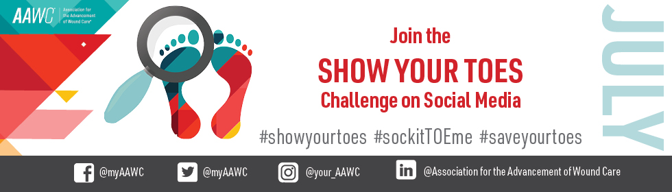
The summer season is upon us, lending good reasons to take off your shoes and SHOW YOUR TOES. It could be taking a walk on a sandy beach or kicking back in a favorite chair by the pool with your feet up. No matter what the reason may be, the AAWC is highlighting a very important reason for you to take your socks and shoes off this month and show your toes

Please JOIN THE AAWC as we celebrate this month by showing your toes through candid photos as you learn more about different arterial and vascular diseases that can be detected through the current condition of your feet. We encourage you, your friends and your family to join this important effort.
Share photos of toes and feet all month long, along with this article to bring awareness to the importance of allowing your doctor to inspect your feet and toes at each visit. Use the following hashtags with each post and share as many photos of as you wish: 
  • #showyourtoes
  • #sockitTOEme 
  • #saveyourtoes
 Use this link if you would like for us to post your photos for you:

Why Show Your Toes?

According to The American Heart Association (AHA), removing one’s socks and shoes to allow the doctor to check the feet for sores, skin color changes and numbness, might just save the limbs or life by detecting issues that are often hidden without this inspection. Underlying problems often present on the feet first. Early detection of abnormal signs leading to disease might help prevent any irreversible conditions. 

Why you need to show your toes:
  1. Peripheral arterial disease (PAD) affects approximately eight and a half million Americans (per the American Heart Association). PAD narrows the arteries to the legs, stomach, arms and head within the upper and lower extremities. This narrowing of the arteries, called atherosclerosis, is caused by plague (fatty deposits) buildup in the arteries which prevents oxygen from moving through the arteries to get to other vital organs, including the skin.

    Symptoms may include cramping, pain or weakness in the legs while walking, climbing stairs or other types of exercise that require more blood flow through the limbs. These symptoms stop when the extra demand from exercise stops or the body is at rest. NOTE: Nearly half of the patients diagnosed with PAD do not show any symptoms, yet show functional impairment when tested. Thus, several people are walking around undiagnosed! 

  2. Other diseases or conditions that can be detected are diabetic foot ulcers and peripheral neuropathy (burning, stinging or numbness of the feet and toes), plus any sensation that makes one feel off balance. A doctor can test for diminished sensation in the feet and toes.

  3. Coronary artery or heart disease is noticeable with skin color and texture changes [skin could become shiny, tight and discolored] thereby helping your doctor diagnose the cause.

    Other signs that arterial narrowing could be present is a lack of hair and nail growth along with atrophy in the muscles of the lower extremities and feet.

  4. Your doctor has many tests that can help evaluate your condition so you can receive treatment that may protect you from progression of disease or relieve symptoms. 
If symptoms are not typical and persist when at rest (called “rest pain”), a doctor may be able to help avoid a more serious issue that could lead to the loss of a limb, so showing your toes is vital!
 
Arterial or vascular disease high-risk factors include high blood pressure, diabetes, or habits such as smoking or poor diets high in fats that elevate cholesterol levels. Other common risk factors include: sex, ethnicity, older age and renal insufficiency; and sometimes pregnancy complications.

Several tests are available to diagnose disease. During an exam, your doctor will perform the following:
  • Check for weak pulses
  • Listen for poor blood flow in the legs
  • Examine and look for any problems on the legs and feet, including hair loss, cold or pale skin and nail growth
Your doctor might also order the following tests/procedures:
  • Blood tests to test cholesterol levels, triglycerides and blood sugar levels
  • Ankle-brachial index test (a vascular test to compare blood pressure in the ankle with the blood pressure in the arm, thereby detecting PAD)
  • Doppler ultrasound to locate those areas with reduced blood flow (blockages) in leg arteries
  • Angiography imaging using contrast dye to specifically locate any blockages

Join the AAWC in helping bring awareness for cardiovascular health and overall quality of life when you SHOW YOUR TOES!

Show Your Toes

On your next annual exam, show your toes. Often, patients overlook, cannot see, or do not recognize a problem. The toes, feet, and legs tell a story about chronic conditions like peripheral vascular disease and Diabetes. Early detection can lead to eliminating preventable amputations.  

According to the American Heart Association, "Peripheral artery disease (PAD) is often used interchangeably with the term, peripheral vascular disease (PVD). PAD stems from structural changes in the blood vessels resulting from fatty buildup (atherosclerosis) in the inner walls of the arteries. These deposits hinder and block normal blood flow."

Peripheral vascular disease (PVD) is an overarching term encompassing vascular diseases resulting from circulatory dysfunction caused by damage to arteries or veins. The most common types of PVD are peripheral artery disease (PAD), chronic venous insufficiency (CVI), and deep vein thrombosis (DVT). There are several signs of vascular complications.

Vascular Disease and The 6P's:
  • Pulselessness - lack of blood flow in the circulation of the large arteries
  • Poikilothermic - decreased skin temperature
  • Pallor - paleness due to a reduced blood supply to the skin
  • Paralysis - the loss of the ability to move a part of the body
  • Paresthesia - burning, prickling, or numb sensation 
  • Pain - no definition needed
Patients and healthcare providers should look for changes in the skin, such as pale or blue toes instead of pink; or increased darkness/pigmentation in dark-skinned patients.  Similarly, thin, brittle, dry, cracked, leathery, shiny, waxy skin on the legs and feet are signs of PVD. Sometimes, a person may start with eczema, a rash, or itchy skin. These may develop due to insufficient blood and oxygen supply to the skin.

Weak pulses or pulselessness in the legs and the feet, including decreased skin temperature, can lead to gangrene, dead tissue due to lack of blood flow. In addition, paresthesia, the loss of sensation to the area, and paralysis, loss of function, usually accompany gangrene. Wounds or sores may develop on the toes, feet, or legs that may bleed, be intensely painful, and slow to heal. Poor blood flow to the skin and surrounding tissue hinders the body's healing ability.

Peripheral arterial disease (PAD) is a complication of diabetes. The prevalence of Diabetes in the US is currently 11.3% of the US, and 38.0% of the adult US population is prediabetic. Furthermore, 48.8% of 65 years or older will be diabetic. Therefore, one person in a senior couple will have Diabetes.  

The signs of diabetes are similar to PVD, but they have the added complication of neuropathy. Neuropathy is the loss of proper sensation, therefore if a person cannot feel pain, they may not know they have a problem or injury. This can cause a delay in their response to seek treatment.

The signs of diabetes-related neuropathy may include:
  • Significant changes to the skin or toenails, including cuts, blisters, calluses, or sores
  • Discharge of fluid/pus or foul-smelling wound
  • Redness and swelling in a joint, like the midfoot or ankle
  • Hyperpigmented or darkened skin on the affected area
  • Corns or calluses are spots of rough skin caused by too much rubbing or pressure in the same spot
  • Warm spots, areas of increased pressure
  • Ingrown toenails
  • Inability to sense hot or cold
  • Burning, numbness & tingling in the feet or legs

Showing your toes needs to be a part of your annual exam, because you may know or not know you have a problem, like PAD or diabetes. More importantly, serious foot problems like these can lead to amputations. Let's work to eliminate preventable amputations.

Please join AAWC during the month of July for our #ShowYourToes challenge! Post a photo of toes to social media with the hashtags #showyourtoes #sockitTOEme #saveyourtoes. Or, submit your photo, and we will post for you to the AAWC channels directly. Thank you for helping to spread awareness of this important issue!
 
For more information on this and related topics, be sure to register for the AAWC Annual Conference November 10-12 in Salt Lake Cityhttps://www.aawconline.org/2022-annual-conference


The Influence of Methamphetamine (Meth) on Wound Healing

Introduction

Because of my son's hardships with meth addiction and assault wounds while incarcerated over the last decade, I decided to be transparent and use my personal experiences and knowledge in the wound care specialty to provide a different perspective. Wound care professionals often highlight how chronic wounds are a public health crisis. However, substance abuse is also a public health crisis that many do not want to discuss. They both have a direct impact on millions of Americans each year. Consider how substance abusers are more likely to develop diabetes, vascular disease, and wound chronicity. We, as healthcare professionals, need to expand our views and our efforts.

Meth-related Wound Challenges 

Meth-related wounds are a significant wound care problem in various clinical care settings. Many nursing facilities around the country refuse to accept such patients, frequently due to stigma, staff training shortages, and liability. Nurse case managers have reported that it is virtually impossible to find placement for these patients. ₃

Additionally, the annual expense of treating meth-associated chronic wounds is a significant financial burden on our healthcare system. While there is a wealth of evidence addressing the behavioral and cognitive impairments produced by meth use in drug users, there is a shortage of information regarding the drug's effect on wound healing and inflammation. ¹ 

Side Effects of Meth 

Methamphetamine (meth) is believed to be used by 35 million people worldwide, including over 10 million users in the United States. Chronic meth addiction and dependence can have several harmful physical and cognitive health consequences. Because the central nervous system contains a high quantity of dopamine, injecting drugs such as meth generates almost immediate euphoric effects. Notably, everyday meth users frequently experience formication, a sensation that feels like insects crawling on or beneath the skin (referred to as meth mites, meth bugs, ice mites, or crank bugs). As a result of formication, users engage in persistent dermotillomania (skin picking, excoriation), resulting in the creation of ulcers that frequently scar. A severe lack of cleanliness among users may also be associated with increased risks of skin infections.

Chronic Conditions in Meth Users

Adults who use methamphetamine are nearly twice as likely to have multiple comorbidities, more than three times as likely to have mental illness, and more than four times as likely to have a substance use disorder, compared to adults who did not. Many people struggle with a combination of medical, mental, and substance use disorders, sometimes all three at the same time. Researchers found that methamphetamine users had a greater prevalence of liver disease (hepatitis or cirrhosis), lung disease (COPD or asthma), and HIV/AIDS among the chronic illnesses evaluated. Additionally, researchers discovered that people who used methamphetamine had a significantly higher risk of developing substance use disorders (SUD) than those who used heroin, prescription stimulants, prescription opioids, cocaine, or sedatives. ₄

Recent Research 

Recent research data indicate that mice treated with meth had a lower ability to repair wounds. Meth stimulates the production of IL-6 in skin tissue, and higher levels of this cytokine may have a direct role in inflammation. Dopamine, which can activate inflammatory cells, may control IL-6. There is a correlation between the effects of leukocyte recruitment and the increase of IL-6 production via dopaminergic activation, thereby aggravating the inflammatory response and prolonging wound healing. There is limited research on the effect of meth use on immunological function, although it appears to have a profound impact on host immunity. ¹ 

Results in How Meth Alters Wound Healing

  • Impairs wound healing by mediating host matrix metalloproteinase-2 collagen degradation²
  • Promotes apoptosis in thymic and splenic lymphocytes when injected 25 mg/kg of body weight into rats¹
  • Decreases thymic and splenic cellularity and affects peripheral T lymphocyte populations in mice¹
  • Damages the mitochondria and causes primary human T cells to malfunction. ¹
  • Increases MRSA burden¹
  • Alters wound healing¹ ²
  • Decreases the number of phagocytic cells in the blood of treated BALB/c mice. ¹ ²
  • Enhances S. aureus biofilm formation²
  • Affects murine neutrophil functions²
  • Impedes the effector functions of human neutrophils²
  • Weakens macrophage function ²
  • Modifies cytokine expression ¹ ²

Link Between Meth Use and MRSA

Although there is a definite clinical correlation between meth use and MRSA disease, there has been no established biological link between increased susceptibility to S. aureus and a meth user's immune response and wound healing capacity. However, animal studies indicate that meth lowers both innate and adaptive immunity and affects the expression of immune cell genes. The purpose of this study was to determine whether meth enhances MRSA skin infections and to demonstrate that the drug has detrimental effects on phagocytic cells generated from humans. ²

Conclusion

Methamphetamine is a significant health concern to our society as it alters people's behavior, putting users at an increased risk of acquiring cutaneous wounds that can become chronic and infected. In the future, the new data will be used to establish realistic treatment options for preventing and managing chronic wounds in drug users. However, it is critical to treat substance abuse, mental illness, and chronic conditions simultaneously.

References

  1. Martinez, L. R. (2019, July 31). Impact of Methamphetamine Induced IL-6 Production on Wound Healing and Inflammation. Grantome NIH. https://grantome.com/grant/NIH/R15-GM117501-01A1
  2. Mihu, M., Roman-Sosa, J., & Varshney, A. (2015, October 7). Methamphetamine Alters the Antimicrobial Efficacy of Phagocytic Cells during Methicillin-Resistant Staphylococcus aureus Skin Infection. mBio, 6(6), 1622-15. doi:10.1128/mBio.01622-15
  3. Bond, A. (2018, April 17). Nursing homes routinely refuse people on addiction treatment. Stat News. Accessed February 10, 2022, from https://www.statnews.com/2018/04/17/nursing-homes-addiction-treatment/
  4. NYU. (2021, Jun 3) Health and Medicine. People Who Use Methamphetamine Likely to Report Multiple Chronic Conditions. https://www.nyu.edu/about/news-publications/news/2021/june/methamphetamine-multiple-chronic-conditions.html. Accessed February 8, 2022. 


When an Obstacle Leads to a Better Solution

Working with the wounded returning from Afghanistan during 2009 through 2011 was extremely challenging.  The wounds I saw during this time were the worst I had seen since arriving at National Naval Medical Center (NNMC), Bethesda in 2007.  The reason was partly due to the mission modification in Afghanistan by the US military in late 2009, when the Marines and Army began to engage in “dismounted” patrols or walking patrols versus patrolling in armored vehicles.  They were exposed and thus more susceptible to injury from improvised explosive devices (IEDs).  Their injuries were likely to be catastrophic because the enemy employed a technique that caused an IED to explode as the Marine/Soldier was standing directly over the device.  The explosions were devastating - these IEDs caused significant soft-tissue injuries and multiple amputations.  The force of the blast drove sand, dirt, debris, and organisms deep into the tissues, causing additional complications.

At NNMC and Walter Reed Army Medical Center (WRAMC) the typical method for managing these catastrophic wounds included serial debridements and washouts in the OR followed by the application of negative pressure wound therapy (NPWT) using reticulated open cell foam dressings (ROCF).  At that time, the trauma teams exclusively used the ROCF dressings containing silver due to their antimicrobial properties.  However, in late 2009 the manufacturer of the ROCF dressings notified us that these dressings would not be available for at least six months.  Because of the complexity of these wounds and the high potential for infection an alternative solution was needed immediately.  We began placing thin layers/sheets of silver impregnated dressings directly in the wound bed and then applying the ROCF dressing on top.  This practice seemed to be effective but not ideal - the application process was more complicated because it was difficult to get good coverage with the silver sheet dressings.

Soon after the silver impregnated ROCF dressings were no longer available we were faced with another issue.  Combat-wounded patients began developing angio-invasive fungal infections (Aspergillus) from the soil imbedded deep in the tissues of their wounds. The next challenge was to implement a therapy that would support NPWT and provide topical antibacterial and antifungal coverage.  Our topical wound therapy included NPWT with the instillation of antimicrobial fluid directly into the wound.  Since this therapy was not frequently being used at this time, it was difficult to find more than a couple of the NPWT units equipped with this function available for use.  The next challenge was to select the appropriate solution as it needed to be antibacterial, antifungal, and non-damaging to healthy wound tissue. 

Our research led us to use a dilute Dakin’s solution of 0.025%, and the initial results were very positive.  Prior to using the Dakin’s instillation with NPWT we would observe black mold growing in the wound as soon as 2-3 days after a debridement and washout.  After initiating the Dakin’s solution as an instillation during NPWT, we no longer saw the black mold within the wound bed.  The treatment was so effective that NPWT with instillation (NPWTi) of dilute Dakin’s became the standard of care for our combat-wounded with complex soft-tissue injuries.  Additionally, the Clinical Practice Guideline for initial management of blast injuries with significant soft-tissue injury and suspected fungal infection was changed to include the use of 0.025% Dakin’s solution moistened gauze as the preferred initial dressing after debridement in the combat theatre.

We typically performed (NPWT) dressing changes 3 times a week during trips to the OR while the patient was undergoing debridements and washouts.  We found that this worked best as the wounds were very large and anesthesia was necessary.  We decreased to twice weekly as the wounds improved and the mold growth decreased.  The surgeons typically used the 0.025% Dakin’s solution until they were ready to perform a skin graft.  This time period varied from 2 to 3 weeks depending on wound progression and tissue cultures.  These wounds were heavily contaminated and often were associated with traumatic amputations.

It is important to see the recent increased attention given to the use of negative pressure wound therapy with instillation in both traumatic and chronic wounds.  The lack of availability of those silver dressings had a far-reaching impact on our care of the combat-wounded as well as future civilian wound care

CDR (Ret) David Crumbley was the Coordinator for the Complex Wound and Limb Salvage program at Walter Reed National Military Medical Center from 2008-2011.  He is currently Professor of Practice at Auburn University School of Nursing and continues to practice wound care at Baptist South Medical Center, Montgomery, Alabama.

Guidelines for Scientific Writing

The art of scientific writing has guidelines that distinguish it from literary writing, documentation, or conversation, especially when preparing a journal article for publication.  Dr. Michael Schneir of the Ostrow School of Dentistry, University of Southern California, is a master of scientific writing and I was fortunate to spend over a year studying with him while preparing my first educational module. I still keep his 600+ page handbook on my desk whenever I write.  At the request of the Communications Committee, here are some of the guidelines that can help avoid the most frequent mistakes I find when editing papers.

Organization

A journal article is organized into the following headings: Abstract, Introduction, Materials and Methods, Results, Discussion, Conclusions or Summary, and References.  Each heading is recommended to have conceptual components with sub-headings that focus on a grouping of information, including a topic sentence and flow of information from general to specific (termed the deductive flow of information).  In producing the article, Dr. Schneir suggests doing the most objective components (e.g. tables, graphs, charts) first, followed by the narrative exposition (results), interpretation of the results (discussion), introduction to the results (introduction), and lastly, the abstract.  Because it is the second most frequently read part of the article (after the title), the abstract needs to be interesting and conceptual with the following components: background information, rationale, purpose, hypothesis, conclusion, and significance.  I remember one graduate school professor stating, “One can learn a lot from reading just the abstract.”  Make every word count!!!

Word Selection

When we had one-on-one sessions over something I had written, Dr. Schneir could spend an hour discussing the option of “a” or “the” before a noun.  For example, is it “a sample” or “the sample”?  “A” is indefinite and singular, whereas “the” is definite and can be either singular or plural and refers to a previously mentioned noun or concept.  Some other words that require careful consideration are which/that (I was often called the “which witch”!), about/concerning, among/between, thereby/thus/because, kind/type – and the list is endless!!

Another concern with word selection is to avoid using literary phrases or words, instead use medical or scientific terminology, e.g. Q-tip in medical terms is cotton-tipped applicator, bed sores are pressure injuries, nowadays is currently.  “Further” implies a distance, whereas “in addition” alerts the reader to another idea relevant to the topic.

The medical community has agreed that providers do not treat diagnoses, they treat patients who have disorders, e.g. patients with diabetes rather than diabetics. 

The use of personal pronouns in scientific writing is discouraged and can usually be avoided by changing the sentence from the active voice (using I, we, they) to the passive voice of the verb.  For example, “we saw the patient in our clinic one week later.”  The passive voice would be “the patient was seen in the clinic one week later.”  Thus, the article reads more scientifically and less like a personal journal.  In addition, words within a sentence are advised to agree in terms of numbers.  For example, after 10 treatment sessions, the patient was discharged to their homes with caretakers to provide daily dressing changes.  The patient has one home, and daily is once per day.  The sentence could be reworded this way: The patient was discharged home with a caretaker instructed to change the dressing daily.  These are very subtle innuendoes, but they become distractors to an experienced reader.

Sentence structure

Whenever possible, it is suggested to combine short, choppy sentences that have some common thread of thought into one sentence that expresses the entire conceptual idea.  For example, the following two sentences can be combined into one: The patient did not have any signs of wound infection.  The team decided not to prescribe any antibiotics for the patient. OR Because there were no signs of wound infection, antibiotics were not indicated.  This also save words and space in the article, and the reader has fewer words to process; therefore the intent is more easily grasped.

Prepositional phrases add unnecessary words to a sentence and can be avoided by transferring the objective of the preposition (a noun) before another noun, thus making it an adjective.  For example, “complications after a stroke” becomes “post-stroke complications.”

Any adjective, adverb, or descriptive phrase should be as close as possible to the noun or verb that is being modified.   And speaking of “should,” the writer does not want to sound authoritative; therefore, words such as advised, recommended, or suggested are similar alternatives that do not leave the reader feeling “preached to” by the author.

Punctuation

The most common mistake observed in punctuation is the use of a colon after “following.”  Colons are not to be used between the verb and its direct objects, but rather after “the following:” and a subsequent list of items.  For example:  Possible causes of lower extremity edema include the following: trauma, chronic venous insufficiency, medications, and congestive heart failure. 

Semicolons are used 1) to separate two independent clauses of a compound sentence, 2) to separate compound elements of a sentence when one or more of the elements contains a comma, and 3) when the elements of a sentence are long. 

Summary

In closing, I will break all of the above rules and share with you a non-scientific pearl of wisdom about writing.  Before ever putting pencil to paper or fingers to keyboard, I close my eyes and dig deep into my soul and ask, what do I really want to tell the reader?  This helps me to organize my thoughts, focus my concepts, and eliminate extraneous information. 

Any of you reading this blog have a story to tell (either a patient case, a clinical dilemma and solution, a pilot study, or organized research) that will help all of us become better clinicians for patients with wounds.  I hope this is helpful, but know that it only scratches the surface of what Dr. Schneir would tell you!!!

1 Comments

The Pressure Is On: Pressure Injury Basics

Pressure Injuries are skin and/or tissue damage which occurs a result of pressure and shear forces which usually occurs over a bony prominence, for example the occiput, cheek bones, chin, shoulders, scapula, spinous process, greater trochanter, sacrum, coccyx, ischium, knee, ankle, and heel. Persons who develop pressure injuries may be chair/bedbound and/or may have some sensory deficit which may not alert the patient about the need to shift positions. Some persons are more at risk to develop a pressure injury due a medical diagnosis, such as a stroke, low or high body mass index, contractures, or a decrease in nutritional intake. By reviewing some of the basics of a pressure injury, formerly known as a pressure ulcer, one may be able to better assess those patients at risk for these skin concerns.

Contrary to popular belief, every incidence of skin breakdown is not a pressure injury. Skin tears present a challenge due to the severity that may be mistaken for a pressure injury. The International Skin Tear Advisory Panel (ISTAP) has established a separate classification for skin tears which notes skin tears as Type 1, Type 2, and Type 3. A Type I skin tears is a tear which has a skin flap which could be replaced to cover the wound.  A Type 2 skin tear has a small skin flap, but the flap does not cover the entire wound.  Type 3 skin tears do not have a skin flap, which causes the entire wound to be visible. Another cause of skin breakdown may be moisture associated skin damage (MASD), which is caused by an overexposure of moisture to the skin and there allows for forces such as friction and shear to increase the risk of pressure injury.  The moisture may be due to urinary or fecal incontinence, wound drainage, fistula output, or perspiration. Over exposure to urinary or fecal incontinence is known as Incontinence Associated Dermatitis or IAD.   Inflammation and skin breakdown related to IAD may be misidentified as a pressure injury wound.

There are unique circumstances when a clinician may question if the wound is related to pressure or another factor. Some examples are:

  1. An immobile person may develop an area of skin breakdown due to moisture associated dermatitis coupled with the evidence of pressure injury to the bony prominence. In such an instance, the bony prominence in question may be palpated to further determine if pressure may be included as a causative factor.
  2. An ambulatory patient has a diagnosis of Diabetes Mellitus and has a full thickness wound to the plantar surface of the foot.  This wound would be considered a diabetic foot wound or a neuropathic wound. Diabetic neuropathy or a decrease in sensation to the foot is the real culprit in this case, in addition to repeated mechanical forces on bony abnormalities.
  3. A paraplegic patient with a diagnosis of Diabetes Mellitus now has full thickness loss to the right heel. Would the wound be considered a neuropathic wound or a pressure injury? In this case, the cause of the wound to the heel would be likely due to pressure.

Another issue which can challenge a clinician is accurately staging a pressure injury.  Only pressure injuries are staged. One would not stage an arterial wound or a surgical wound. Terms such as a partial thickness or full thickness tissue loss are used in the description of these wounds, but they would not be given a stage. Once a pressure injury is identified as a certain stage, the injury is not “back staged”. For example:

From the onset, the wound is noted to be a stage 3 pressure injury.  As the wound begins to heal, it will be noted as a stage 3 pressure injury in the proliferative or remodeling phase of healing. The wound will not progress from a stage 3, then to a stage 2 or to a stage 1 pressure injury.

Pressure Injuries Stages at a Glance:

Stage 1: Intact skin over a bony prominence with erythema that does not blanch to the touch.  Key word is INTACT. In some individuals, this site resembles a sunburn.  In darker skin persons, the site in question may not have evidence of erythema. The patient may not have an increase in pain to the site. However, the clinician may notice a difference in temperature, texture, or hardness to the area of concern and the surrounding skin when palpated.

Stage 2: Partial thickness loss usually over a bony prominence. The tissue loss extends to the dermis, but no subcutaneous tissue is observed. Blisters that contain serous or clear fluid are also defined as Stage 2. 

Stage 3: Full thickness tissue loss usually over a bony prominence. The tissue loss extends to the subcutaneous tissue. Slough, tunneling, or undermining may be noted, but no tendon, bone, or muscle is noted.

Stage 4: Full thickness tissue loss over a bony prominence with visible tendon, bone, or muscle. If cartilage is visible to areas such as the nose or ear, the wound is considered a Stage 4 pressure injury.

Unstageable pressure injuries are mostly covered by slough or eschar. The amount of tissue loss is unknown because the depth of the wound is unseen.

Device related pressure injuries are injuries caused by a medical device, such as a nasal cannula or ill-fitting thromboembolic hose.

If a wound is noted in the mouth or mucous membranes, list this wound as a mucosal injury, but do not stage this injury as a pressure wound.

In a wound with Deep Tissue Pressure Injury (DTPI), the skin is intact or there may be an intact blister that appears maroon or purple due to the deep tissue bleeding that has occurred due to the capillary disruption in the deep tissue. These wounds may evolve into a full thickness wound and become a Stage 3 or Stage 4 pressure injury.

Now that pressure injuries have been identified, as well as a few other skin concerns, the goal is to identify those patients at risk, as well as endeavor to prevent and manage these life changing skin issues for the patient! 


References:

Beeckman D, Campbell J, Campbell K et al. (2015).  Incontinence associated dermatitis: moving prevention forward. Proceedings of the Global IAD Expert Panel. Wounds International. https://tinyurl.com/ycmyfv2d.

FISHER, P., & HIMAN, C. (2020). Moisture-associated skin damage: a skin issue more prevalent than pressure ulcers. Wounds UK16(1), 58–63.

Gray, M. , Black, J. M. , Baharestani, M. M. , Bliss, D. Z. , Colwell, J. C. , Goldberg, M. , Kennedy-Evans, K. L. , Logan, S. & Ratliff, C. R. (2011). Moisture-Associated Skin Damage. Journal of Wound, Ostomy and Continence Nursing, 38(3), 233–241.  doi:10.1097/WON.0b013e318215f798.

International Skin Tear Advisory Panel (ISTAP) (n.d.). ISTAP skin tear classification. Retrieved March 15, 2021 from http://www.skintears.org/education/tools/istap-skin-tear-classification/

LeBlanc, K., Alam, T., Langemo, D., Baranoski, S., Campbell, K., & Woo, K. (2016). Clinical challenges of differentiating skin tears from pressure ulcers. EWMA Journal16(1), 17–23. 

National Pressure Injury Advisory Panel (NPIAP) (n.d.). Pressure injury stages. Retrieved March 15, 2021 from https://npiap.com/page/PressureInjuryStages

National Pressure Injury Advisory Panel (NPIAP) (2017). National Pressure Ulcer Position Statement on Staging-2017 Clarifications. Retrieved March 16, 2021 from https://cdn.ymaws.com/npiap.com/resource/resmgr/npuap-position-statement-on-.pdf

1 Comments

How Does a Whole-Food Plant-Based (WFPB) Diet Work in Wound Care? Seven WFPB nutrition tips to remember in wound care.

Today, we will discuss seven WFPB nutrition tips that apply to wound care and the role of the wound care physician/practitioner in caring for a patient with a disease that can be complicated by poor diet and lifestyle habits. While reading today, take the time to reflect on what obligations we have toward those who are at risk but have not sought our help. Any wound care practice must be evidence-based. Just as prescribing services must be founded on solid evidence, the same is true for dietary advice.  Was there a sincere evaluation of the patient's nutrition status? What should be done next with this information? Should time be spent reviewing articles to place in your personal armamentarium?  Primary care physicians do not perform general surgeries or run  urinalysis; neither do they need to do their own diet counseling. That is the job of a qualified dietitian/nutritionist. The wound care practitioner simply needs to know that nutrition is important, wound healing nutritional deficiencies need correction, and clear communication with the patient is imperative, while providing a solid referral to other interdisciplinary professionals.

TIP #1: The building evidence of a healthy diet
What type of diet is recommended for a patient with a wound?
With accumulative scientific evidence supporting a healthy diet, whole-food plant-based (WFPB) diets are steadily on the rise. A WFPB diet consists of vegetables, fruits, legumes, whole grains, nuts, seeds, herbs, and spices. The United States Department of Agriculture, American Heart Association, American Institute for Cancer Research, National Kidney Foundation, and American Diabetes Association all promote WFPB diets, and it doesn't have to be an all or nothing.  Evidence-based studies show a positive correlation between WFPB diet compliance and overall well-being.

TIP #2: WFPB benefits
Why are WFPB diets becoming more popular? 
Aside from improved healing with proper nutrition, what benefits are there to my patient? WFPB diets are associated with:

  1. Lowering overall mortality
  2. Lowering ischemic heart disease mortality
  3. Supporting a healthy weight
  4. Reducing medications
  5. Reducing obesity
  6. Reducing obesity-related inflammatory markers
  7. Reducing hyperglycemia
  8. Reducing hyperlipidemia
  9. Reducing hypertension
  10. Reversing advanced cardiovascular disease
  11. Reversing Type 2 Diabetes.

TIP #3: Protein
Can the protein needs of wound and geriatric patients be met with WFPB diets? 
Absolutely! The primary food sources for proteins are legumes (beans, lentils, peas, peanuts), nuts, seeds, and soy foods. Poor appetite and poor dentition are complications with geriatric wound care patients, making meat alternatives, such as smoothies, cooked cereals, lentil or bean-rich soups, and spreadable hummus nutritionally dense additions to their diets. Nevertheless, one must remember to temper the increased desire for a high protein wound care diet and the need to satisfy a low protein diet in chronic kidney disease patients, especially CKD Stage II and Stage III. This is similar to the scenario of a patient with severe lymphedema, aggressive congestive heart failure, or pulmonary edema.

**Brenner BM. Remission of renal disease: recounting the challenge, acquiring the goal. J Clin Invest. 2002;110(12):1753-1758. doi:10.1172/JCI17351

TIP #4: Vitamin B12
Will you have to supplement vitamin B12? 
Yes! Numerous research studies demonstrate vitamin B12 supplementation in metformin-treated type 2 diabetes patients is beneficial in order to prevent the occurrence of vitamin B12 deficiency complications.  Deficiency in the elderly population is high due to inadequate intake and malabsorption. Treatments are safe and effective for adults over 60 years, regardless of their diet. Vitamin D should also be supplemented if sun exposure is not a viable option for patients.

**Goraya N, Munoz-Maldonado Y, Simoni J, Wesson DE. Fruit and Vegetable Treatment of Chronic Kidney Disease-Related Metabolic Acidosis Reduces Cardiovascular Risk Better than Sodium Bicarbonate. Am J Nephrol. 2019;49(6):438-448. doi:10.1159/000500042


TIP #5: Nutritional Drinks
Are commercial products the only option for complete nutrition, muscle loss prevention, and wound care healing? No. Therapeutic nutrition drinks and powders have been shown to support wound healing clinically. However, eating a balanced diet high in fiber and low in animal proteins has shown to support wound healing.  For example, pumpkin seeds have one of the highest concentrations of arginine. Other foods are sesame & sunflower seeds, and tree nuts, which all have high omega-3 fatty acids. Another supplement frequently added to nutritional drinks is beta-hydroxy-beta-methyl butyrate (HMB). But is it necessary?  HMB on exercise performance and body composition did not make a difference when comparing whey, soy, or leucine-enriched soy protein. HMB helps with slowing age-related muscle loss (sarcopenia). How do you balance supplements versus instructing patients to eat healthier with a soy product?

**Wilson GJ, Wilson JM, Manninen AH. Effects of beta-hydroxy-beta-methylbutyrate (HMB) on exercise performance and body composition across varying levels of age, sex, and training experience: A review. Nutr Metab (Lond). 2008;5:1. Published 2008 Jan 3. doi:10.1186/1743-7075-5-1


TIP #6: What about fats? 
A high-fatty diet and alcohol consumption delay the healing process by decreasing stimulation of collagen synthesis and reduction of granulation tissue and reepithelialization. Diabesity is the worldwide twin epidemics of obesity and diabetes. The American Heart Association recommends limiting saturated fats to less than 14 grams in a 2000 calorie daily diet. By the way, trans fats have been banned in the United States since 2018, with an extended compliance date for these foods until January 1, 2020. 

Monounsaturated fat (MUFAs) and polyunsaturated fats (PUFAs) are associated with lowering blood pressure, improving blood cholesterol levels, and decreasing the risk of heart attack and stroke.  Omega-6 PUFAs have a particular role in the structural integrity and barrier function of the skin. Omega-3 PUFAs give aid in signaling molecules that influence the inflammatory response in the skin, while MUFAs aid in angiogenesis and aid in the regulation of insulin levels and blood sugar control. 

Eating a colorful variety of WFPB foods promotes a large variety of micronutrient exposure, antioxidants, and other cellular regulatory properties such as vitamin C, calcium, iron, vitamin K, selenium, zinc, and healthy MUFAs & PUFAs. Nothing new here.  


TIP #7: Got a fiber gap?
Currently, only 5% of the United States population meets the daily target of 25 grams of fiber for women and 38 grams of fiber for men. The benefits of prescribing a high fiber diet are correlated with a reduction in comorbidities for patients with wounds. However, how does a high fiber diet help wound care patients? Well, gut bacteria of vegans produce neuroactive molecules like gamma-aminobutyric acid (GABA) which in turnreduces the stress response in humans, along with decreasing cortisol levels. GABA also counters high glucocorticoid levels, which impair wound healing. Overall, WFPB high fiber diets modulate GABA, which regulates blood pressure and heart rate, influences GI motility, and plays a role in anxiety, depression, pain sensation, and immune response.  Emotionally happy wound patients with reduced stress heal faster. 

**Briguglio M, Dell'Osso B, Panzica G, et al. Dietary Neurotransmitters: A Narrative Review on Current Knowledge. Nutrients. 2018;10(5):591. Published 2018 May 10. doi:10.3390/nu10050591

**Ebrecht M, Hextall J, Kirtley LG, Taylor A, Dyson M, Weinman J. Perceived stress and cortisol levels predict speed of wound healing in healthy male adults. Psychoneuroendocrinology. 2004;29(6):798-809. doi:10.1016/S0306-4530(03)00144-6


Finally, the wrap-up,  I hope this stimulates your view into the world of nutrition, WFPB, and patient care. Jump into your research and become a student in the adventure toward better patient care. None of the information is new, but the POV might be. History has taught us that Occam's razor or law of parsimony states, "plurality should not be posited without necessity" --  translation -- The state of fact should not be assumed as a fact.

It's all about the simplicity of two competing theories; the simpler explanation of an entity is to be preferred. 

Unhealthful eating habits affect the whole family. Medications are no substitute for dietary interventions.

 
2 Comments